| Info
Sheets |
| | | | | | | | | | | | | | | | | | | | | | | | |
 | Out-
side |
| | | | |
|
| | | | |
Result : Searchterm 'high field' found in 1 term [ ] and 35 definitions [ ] and 35 definitions [ ] ]
| | previous 16 - 20 (of 36) nextResult Pages : [1] [2 3 4 5 6 7 8] |  | |  | Searchterm 'high field' was also found in the following services: | | | | |
| |  |
| |
|

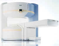
From Philips Medical Systems;
Philips continues to expand the frontiers of utra high field MRI with the introduction of the new Intera Achieva 3.0T™. Its powerful future-safe platform shares all the advantages of the Achieva family and covers applications throughout the whole body.
Device Information and Specification
CLINICAL APPLICATION
Whole body
CONFIGURATION
Short bore compact
SE, Modified-SE, IR (T1, T2, PD), STIR, FLAIR, SPIR, FFE, T1-FFE, T2-FFE, Balanced FFE, TFE, Balanced TFE, Dynamic, Keyhole, 3D, Multi Chunk 3D, Multi Stack 3D, K Space Shutter, MTC, TSE, Dual IR, DRIVE, EPI, Cine, 2DMSS, DAVE, Mixed Mode; Angiography: Inflow MRA, TONE, PCA, CE MRA
128 x 128, 256 x 256,512 x 512,1024 x 1024 (64 for Bold img)
Variable in 1% increments
Lum.: 120 cd/m2; contrast: 150:1
Variable (op. param. depend.)
POWER REQUIREMENTS
380/400 V
Passive and dynamic, 1st order std./2nd opt.
| | | | | | | | | | |  Further Reading: Further Reading: | News & More:
|
|
| |
| | | | | |
| |
|
Knee MRI, with its high soft tissue contrast is one of the main imaging tools to depict knee joint pathology. MRI allows accurate imaging of intra-articular structures such as ligaments, cartilage, menisci, bone marrow, synovium, and adjacent soft tissue.
Knee exams require a dedicated extremity coil, providing a homogenous imaging volume and high SNR to ensure best signal coverage.
A complete knee MR examination includes for example sagittal and coronal T1 weighted, and proton density weighted pulse sequences +/- fat saturation, or STIR sequences. For high spatial resolution, maximal 4 mm thick slices with at least an in plane resolution of 0.75 mm and small gap are recommended. To depict the anterior cruciate ligament clearly, the sagittal plane has to be rotated 10 - 20° externally (parallel to the medial border of the femoral condyle). Retropatellar cartilage can bee seen for example in axial T2 weighted gradient echo sequences with Fatsat. However, the choice of the pulse sequences is depended of the diagnostic question, the used scanner, and preference of the operator.
Diagnostic quality in knee imaging is possible with field strengths ranging from 0.2 to 3T. With low field strengths more signal averages must be measured, resulting in increased scan times to provide equivalent quality as high field strengths.
More diagnostic information of meniscal tears and chondral defects can be obtained by direct magnetic resonance arthrography, which is done by introducing a dilute solution of gadolinium in saline (1:1000) into the joint capsule. The knee is then scanned in all three planes using T1W sequences with fat suppression. For indirect arthrography, the contrast is given i.v. and similar scans are started 20 min. after injection and exercise of the knee.
Frequent indications of MRI scans in musculoskeletal knee diseases are: e.g., meniscal degeneration and tears, ligament injuries, osteochondral fractures, osteochondritis dissecans, avascular bone necrosis and rheumatoid arthritis. See also Imaging of the Extremities and STIR. | | | | | |
• View the DATABASE results for 'Knee MRI' (4).
| | |
• View the NEWS results for 'Knee MRI' (4).
| | | | | | Further Reading: | | Basics:
|
|
News & More:
| |
| |
| | | | | |
| |
|
| | | | | | | |
• View the DATABASE results for 'Lung Imaging' (7).
| | |
• View the NEWS results for 'Lung Imaging' (3).
| | | | | | Further Reading: | | Basics:
|
|
News & More:
|  |
Chest MRI a viable alternative to chest CT in COVID-19 pneumonia follow-up
Monday, 21 September 2020 by www.healthimaging.com | | |
CT Imaging Features of 2019 Novel Corona virus (2019-nCoV)
Tuesday, 4 February 2020 by pubs.rsna.org | | |
Polarean Imaging Phase III Trial Results Point to Potential Improvements in Lung Imaging
Wednesday, 29 January 2020 by www.diagnosticimaging.com | | |
Low Power MRI Helps Image Lungs, Brings Costs Down
Thursday, 10 October 2019 by www.medgadget.com | | |
Chest MRI Using Multivane-XD, a Novel T2-Weighted Free Breathing MR Sequence
Thursday, 11 July 2019 by www.sciencedirect.co | | |
Researchers Review Importance of Non-Invasive Imaging in Diagnosis and Management of PAH
Wednesday, 11 March 2015 by lungdiseasenews.com | | |
New MRI Approach Reveals Bronchiectasis' Key Features Within the Lung
Thursday, 13 November 2014 by lungdiseasenews.com | | |
MRI techniques improve pulmonary embolism detection
Monday, 19 March 2012 by medicalxpress.com |
|
News & More:
| |
| |
| | | Searchterm 'high field' was also found in the following services: | | | | |
| | |
| |
|
Device Information and Specification
CLINICAL APPLICATION
Whole body
GRE, IR, FIR, STIR, TrueIR/FISP, FSE, MT, SS-FSE, MT-SE, MTC, MSE, GMR, fat/water sat./exc.
IMAGING MODES
Single, multislice, volume study, multi angle
512 x 512 full screen display
POWER REQUIREMENTS
380/400/420/440/480 V
| | | |
• View the DATABASE results for 'MAGNETOM Concerto™' (2).
| | | | |
| | | | | |
| |
|
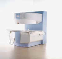
From Siemens Medical Systems;
the MAGNETOM Rhapsody™. This open MRI system offers the proven image
quality of 1.0 Tesla. In addition to the resulting broad range of applications, the open magnet of the high field system MAGNETOM Rhapsodyâ„¢ facilitates examination of claustrophobic and pediatric patients. And the system allows for expanded interventional applications.
Device Information and Specification CLINICAL APPLICATION Whole body GRE, IR, FIR, STIR, TrueIR/FISP, FSE, FLAIR, MT, SS-FSE, MT-SE, MTC, MSE, EPI, GMR, fat/water sat./exc. IMAGING MODES Single, multislice, volume study, multi angle, multi oblique1024 x 1024 full screen display POWER REQUIREMENTS 380/400/420/440/480 V | | | |
• View the DATABASE results for 'MAGNETOM Rhapsody™' (2).
| | | | |
| | | | |
| | | |
|
| |
 | Look
Ups |
| |