 | Info
Sheets |
| | | | | | | | | | | | | | | | | | | | | | | | |
 | Out-
side |
| | | | |
|
| | | | |
Result : Searchterm '3 Tesla' found in 0 term [ ] and 11 definitions [ ] and 11 definitions [ ], (+ 11 Boolean[ ], (+ 11 Boolean[ ] results ] results
| | 1 - 5 (of 22) nextResult Pages : [1 2 3] [4 5] |  | |  | Searchterm '3 Tesla' was also found in the following services: | | | | |
| |  |
| |
|
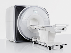
From Siemens Medical Systems;
Received FDA clearance in 2013.
The MAGNETOM Prisma is the 3T PowerPack for exploration that offers most demanding clinical and research challenges of today and the future. The latest parallel transmit technology, TimTX TrueShape, enables zooming into specific body regions for enhanced image quality. Furthermore, the Tim 4G integrated coil technology offers remarkable imaging flexibility and supports complex
examinations across the whole body.
Onsite upgrades to the MAGNETOM Prisma for customers who have already installed the 3 Tesla MAGNETOM Trio are possible.
Device Information and Specification
CLINICAL APPLICATION
Whole Body
CONFIGURATION
Ultra-short bore
Head, spine, torso/ body coil, neurovascular, cardiac, neck, shoulder, knee, wrist, foot//ankle and multi-purpose flex coils. Peripheral vascular, breast, shoulder.
CHANNELS (min. / max. configuration)
64, 128
MAGNET WEIGHT (gantry included)
13000 kg
DIMENSION H*W*D (gantry included)
173 x 230 x 222 cm
Passive, active; first order,
second order
POWER REQUIREMENTS
380 / 400 / 420 / 440 / 460 / 480 V, 3-phase + ground;
| | | | | | • Share the entry 'MAGNETOM Prisma':    | | | | |
 |
| |
|
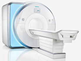
From Siemens Medical Systems;
Received FDA clearance in 2010.
MAGNETOM Skyra is a top-of-the-line, patient friendly wide bore 3 Tesla MRI system.
The system is equipped with the Tim 4G and Dot system (Total imaging matrix and Day optimizing throughput), to enhance both productivity and image quality with the complete range of advanced applications for clinical routine and research. Tim 4G features lighter, trimmer MRI coils that take up less space inside the magnet but deliver a high coil element density with increased signal to noise ratio and the possibility to use high iPAT factors.
Device Information and Specification
CLINICAL APPLICATION
Whole Body
Head, spine, torso/ body coil, neurovascular, cardiac, neck, shoulder, knee, wrist, foot//ankle and multi-purpose flex coils. Peripheral vascular, breast, shoulder.
CHANNELS (min. / max. configuration)
48, 64, 128
Chemical shift imaging, single voxel spectroscopy
MINIMUM TE
3D T1 spoiled GRE: 0.22 (256 matrix), Ultra-short TE
At isocenter: L-R 70 cm, A-P (with table) 55 cm
MAGNET WEIGHT (gantry included)
5768 kg
DIMENSION H*W*D (gantry included)
173 x 231 x 219 cm
COOLING SYSTEM
Water; single cryogen, 2 stage refrigeration
3 linear with 20 coils, 5 nonlinear 2nd-order
POWER REQUIREMENTS
380 / 400 / 420 / 440 / 460 / 480 V, 3-phase + ground; 110 kVA
| | | | | |
| | | | | |
| |
|
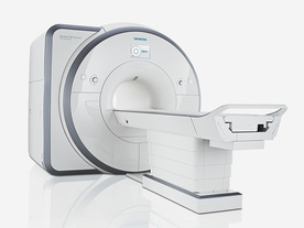
From Siemens Medical Systems;
Received FDA clearance in 2012.
The MAGNETOM Spectra is a cost-optimized high field MRI system with Tim 4G and Dot technologies. The system consumes less energy compared to other 3 Tesla scanners. The magnet-cooling helium is contained in a closed loop, which prevents the gas from escaping and reduces the need for refills. TimTX includes innovative techniques in the radio frequency excitation hardware as well as new application and processing features enabling uniform RF distribution in all body regions.
Device Information and Specification
CLINICAL APPLICATION
Whole Body
Head, spine, torso/ body coil, neurovascular, neck and multi-purpose flex coils. Peripheral vascular, breast, shoulder, knee, wrist, foot//ankle, endorectal optional.
Chemical shift imaging, single voxel spectroscopy
DIMENSION H*W*D (gantry included)
173 x 231 x 219 cm
COOLING SYSTEM
Water; single cryogen, 2 stage refrigeration
Passive, active; first order standard, second order optional
POWER REQUIREMENTS
380 / 400 / 420 / 440 / 460 / 480 V, 3-phase + ground; connection value with chiller 100 kvA /without chiller 60 kVA
| | | | | |
| | | Searchterm '3 Tesla' was also found in the following services: | | | | |
| | |
| |
|

FDA cleared and CE Mark 2011.
The Biograph mMR has a fully-integrated design for simultaneous PET/MRI imaging. The dedicated hardware includes solid-state, avalanche photodiode PET detector and adapted, PET-compatible MR coils.
The possibility of truly simultaneous operation allows the acquisition of several magnetic resonance imaging ( MRI) sequences during the positron emission tomography (PET) scan, without increasing the examination time.
See also Hybrid Imaging.
Device Information and Specification
CLINICAL APPLICATION
Whole Body
CONFIGURATION
Simultaneous PET/MRI
26 cm (typical overlap 23%)
A-P 45, R-L 50, H-F 50 cm
PET RING DIAMETER
65.6 cm
PATIENT SCAN RANGE
199 cm
HORIZONTAL SPEED
200 mmsec
PET DETECTOR
Solid state, 4032 avalanche photo diodes
DETECTOR SCINTILLATION MATERIAL
LSO, 28672 crystals
CRYSTAL SIZE
4 x 4 x 20 mm
DIMENSION H*W*D (gantry included)
335 x 230 x 242 cm (finshed covers)
COOLING SYSTEM
PET system: water; MRI system: water
Aautomatic, patient specific shim; active shim 3 linear and 5 non-linear channels (seond order)
POWER REQUIREMENTS
380 / 400 / 420 / 440 / 460 / 480 V, 3-phase + ground; Total system 110kW
| | | | | |  Further Reading: Further Reading: | | Basics:
|
|
News & More:
| |
| |
| | | | | |
| |
|
The principal advantage of MRI at high field is the increase in signal to noise ratio. This can be used to improve anatomic and/or temporal resolution and reduce scan time while preserving image quality. MRI devices for whole body imaging for human use are available up to 3 tesla (3T). Functional MRI ( fMRI) and MR spectroscopy ( MRS) benefit significantly. In addition, 3T machines have a great utility in applications such as TOF MRA and DTI. Higher field strengths are used for imaging of small parts of the body or scientific animal experiments. Higher contrast may permit reduction of gadolinium doses and, in some cases, earlier detection of disease.
Using high field MRI//MRS, the RF-wavelength and the dimension of the human body complicating the development of MR coils. The absorption of RF power causes heating of the tissue. The energy deposited in the patient's tissues is fourfold higher at 3T than at 1.5T. The specific absorption rate (SAR) induced temperature changes of the human body are the most important safety issue of high field MRI//MRS.
Susceptibility and chemical shift dispersion increase like T1, therefore high field MRI occasionally exhibits imaging artifacts. Most are obvious and easily recognized but some are subtle and mimic diseases. A thorough understanding of these artifacts is important to avoid potential pitfalls. Some imaging techniques or procedures can be utilized to remove or identify artifacts. See also Diffusion Tensor Imaging.
See also the related poll result: ' In 2010 your scanner will probably work with a field strength of' | | | | | |
• View the DATABASE results for 'High Field MRI' (16).
| | |
• View the NEWS results for 'High Field MRI' (9).
| | | | | | Further Reading: | | Basics:
|
|
News & More:
| |
| |
| | | | |
| | | 1 - 5 (of 22) nextResult Pages : [1 2 3] [4 5] |
| |
|
| |
 | Look
Ups |
| |


