The
MRI device is located within a specially
shielded room (
Faraday cage) to avoid outside
interference, caused by the use of radio waves very close in
frequency to those of ordinary FM radio stations.
The
MRI procedure can easily be performed through clothing and bones, but attention must be paid to
ferromagnetic items, because they will be attracted from the
magnetic field. A hospital gown is appropriate, or the patient should wear clothing without metal fasteners and remove any metallic objects like hairpins, jewelry, eyeglasses, clocks, hearing aids, any removable
dental work, lighters, coins etc., not only for
MRI safety reasons.
Metal in or around the scanned area can also cause errors in the reconstructed images (
artifacts). Because the strong
magnetic field can displace, or disrupt metallic objects, people with an implanted
active device like a
cardiac pacemaker cannot be scanned under normal circumstances and should not enter the
MRI area.
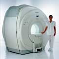
The
MRI machine can look like a short tunnel or has an
open MRI design and the
magnet does not completely surround the patient. Usually the patient lies on a comfortable motorized table, which slides into the scanner, depending on the
MRI device, patients may be also able to sit up. If a
contrast agent is to be administered, intravenous access will be placed. A technologist will operate the
MRI machine and observe the patient during the examination from an adjacent room. Several sets of images are usually required, each taking some minutes. A typical
MRI scan includes three to nine imaging
sequences and may take up to one hour. Improved
MRI devices with powerful magnets, newer software, and advanced
sequences may complete the process in less time and better
image quality.
Before and after the most
MRI procedures no special preparation, diet, reduced activity, and extra medication is necessary. The
magnetic field and radio waves are not felt and no pain is to expect.
Movement can blur
MRI images and cause certain artifacts. A possible problem is the
claustrophobia that some patients experience from being inside a tunnel-like scanner. If someone is very anxious or has difficulty to lie still, a sedative agent may be given. Earplugs and/or headphones are usually given to the patient to reduce the loud
acoustic noise, which the machine produces during normal operation. A technologist observes the patient during the test. Some
MRI scanners are equipped with televisions and music to help the examination time pass.
MRI is not a cheap examination, however cost effective by eliminating the need for invasive radiographic procedures, biopsies, and exploratory surgery.
MRI scans can also save money while minimizing patient risk and discomfort. For example,
MRI can reduce the need for X-ray
angiography and myelography, and can eliminate unnecessary diagnostic procedures that miss occult disease.
See also
Magnetic Resonance Imaging MRI,
Medical Imaging,
Cervical Spine MRI,
Claustrophobia,
MRI Risks and
Pregnancy.
For
Ultrasound Imaging (USI) see
Ultrasound Imaging Procedures at
Medical-Ultrasound-Imaging.com.
See also the related poll result: '
MRI will have replaced 50% of x-ray exams by'


 (GRE - sequence) A