| Info
Sheets |
| | | | | | | | | | | | | | | | | | | | | | | | |
 | Out-
side |
| | | | |
|
| | | | |
Result : Searchterm 'Real Time' found in 2 terms [ ] and 5 definitions [ ] and 5 definitions [ ], (+ 13 Boolean[ ], (+ 13 Boolean[ ] results ] results
| | previous 11 - 15 (of 20) nextResult Pages : [1] [2] [3 4] |  | |  | Searchterm 'Real Time' was also found in the following services: | | | | |
| |  |
| |
|

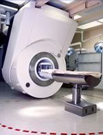
From Philips Medical Systems;
the Panorama 0.23 T, providing a new design optimized for patient comfort, faster reconstruction time than before (300 images/second) and new gradient
specifications. Philips' Panorama 0.23 T I/T supports MR-guided interventions, resulting in minimally invasive procedures, more targeted surgery, reduced recovery time and shorter hospital stays. Optional OptoGuide functionality enables real- time needle tracking. Philips' Panorama 0.23 TPanorama 0.2 R/T is the first and only open MRI system to enable radiation therapy planning using MR data sets. The Panorama also features the new and consistent Philips User Interface, an essential element of the Vequion clinical IT family of products and services.
Device Information and Specification CLINICAL APPLICATION Whole body SE, FE, IR, FFE, DEFFE, DESE, TSE, DETSE, Single shot SE, DRIVE, Balanced FFE, MRCP, Fluid Attenuated Inversion Recovery, Turbo FLAIR, IR-TSE, T1-STIR TSE, T2-STIR TSE, Diffusion Imaging, 3D SE, 3D FFE, MTC;; Angiography: CE-ANGIO, MRA 2D, 3D TOFOpen x 46 cm x infinite (side-first patient entry) POWER REQUIREMENTS 400/480 V COOLING SYSTEM TYPE Closed loop chilled water ( chiller included) | | | | | |  Further Reading: Further Reading: | News & More:
|
|
| |
| | | Searchterm 'Real Time' was also found in the following service: | | | | |
| | |
| |
|
Device Information and Specification
CLINICAL APPLICATION
Whole body
CONFIGURATION
Mobile compact
Whole body, intra-operative head, neck volume, atlas head//neck vascular quadrature phased array, spine quadrature, C/T/L spine phased array, small joint, large joint, TMJ bilateral, shoulder phased array, extremity quadrature volume, wrist, hand quadrature, general purpose flexible, pelvis/abdomen phased array, body quadrature, phased array flexible, breast bilateral
IMAGING MODES
Localizer, single slice, multislice, volume
| | | |
• View the DATABASE results for 'iMotion™ 1.5 Tesla Magnet' (2).
| | | | |
| | | | | |
| |
|

It is important to remember when working around a superconducting magnet that the magnetic field is always on. Under usual working conditions the field is never turned off. Attention must be paid to keep all ferromagnetic items at an adequate distance from the magnet. Ferromagnetic objects which came accidentally under the influence of these strong magnets can injure or kill individuals in or nearby the magnet, or can seriously damage every hardware, the magnet itself, the cooling system, etc..
See MRI resources Accidents.
The doors leading to a magnet room should be closed at all times except when entering or exiting the room. Every person working in or entering the magnet room or adjacent rooms with a magnetic field has to be instructed about the dangers. This should include the patient, intensive-care staff, and maintenance-, service- and cleaning personnel, etc..
The 5 Gauss limit defines the 'safe' level of static magnetic field exposure. The value of the absorbed dose is fixed by the authorities to avoid heating of the patient's tissue and is defined by the specific absorption rate.
Leads or wires that are used in the magnet bore during imaging procedures, should not form large-radius wire loops. Leg-to-leg and leg-to-arm skin contact should be prevented in order to avoid the risk of burning due to the generation of high current loops if the legs or arms are allowed to touch. The patient's skin should not be in contact with the inner bore of the magnet.
The outflow from cryogens like liquid helium is improbable during normal operation and not a real danger for patients.
The safety of MRI contrast agents is tested in drug trials and they have a high compatibility with very few side effects. The variations of the side effects and possible contraindications are similar to X-ray contrast medium, but very rare. In general, an adverse reaction increases with the quantity of the MRI contrast medium and also with the osmolarity of the compound.
See also 5 Gauss Fringe Field, 5 Gauss Line, Cardiac Risks, Cardiac Stent, dB/dt, Legal Requirements, Low Field MRI, Magnetohydrodynamic Effect, MR Compatibility, MR Guided Interventions, Claustrophobia, MRI Risks and Shielding. | | | | | | | | |
• View the DATABASE results for 'MRI Safety' (42).
| | |
• View the NEWS results for 'MRI Safety' (13).
| | | | | | Further Reading: | | Basics:
|
|
News & More:
| |
| |
| | | Searchterm 'Real Time' was also found in the following services: | | | | |
| | |
| |
|
(DANTE) A technique used to place a saturation band over e.g. the myocardium. This technique includes spatial modulation of magnetization complementary and delays alternating with nutations for tailored excitation, followed by the application of a cine or real- time imaging. Because the saturated magnetization pattern moves with the atoms of the tissue, the cardiac motion shows up as deformations in the grid pattern in the resulting imaging sequence. | | | | | |
| | | Searchterm 'Real Time' was also found in the following service: | | | | |
| | |
| |
|
(MRS / MRSI - Magnetic Resonance Spectroscopic Imaging) A method using the NMR phenomenon to identify the chemical state of various elements without destroying the sample. MRS therefore provides information about the chemical composition of the tissues and the changes in chemical composition, which may occur with disease processes.
Although MRS is primarily employed as a research tool and has yet to achieve widespread acceptance in routine clinical practice, there is a growing realization that a noninvasive technique, which monitors disease biochemistry can provide important new information for the clinician.
The underlying principle of MRS is that atomic nuclei are surrounded by a cloud of electrons, which very slightly shield the nucleus from any external magnetic field. As the structure of the electron cloud is specific to an individual molecule or compound, then the magnitude of this screening effect is also a characteristic of the chemical environment of individual nuclei.
In view of the fact that the resonant frequency is proportional to the magnetic field that it experiences, it follows that the resonant frequency will be determined not only by the external applied field, but also by the small field shift generated by the electron cloud.
This shift in frequency is called the chemical shift (see also Chemical Shift). It should be noted that chemical shift is a very small effect, usually expressed in ppm of the main frequency. In order to resolve the different chemical species, it is therefore necessary to achieve very high levels of homogeneity of the main magnetic field B0.
Spectra from humans usually require shimming the magnet to approximately one part in 100. High resolution spectra of liquid samples demand a homogeneity of about one part in 1000.
In addition to the effects of factors such as relaxation times that can affect the NMR signal, as seen in magnetic resonance imaging, effects such as J-modulation or the transfer of magnetization after selective excitation of particular spectral lines can affect the relative strengths of spectral lines.
In the context of human MRS, two nuclei are of particular interest - H-1 and P-31. (PMRS - Proton Magnetic Resonance Spectroscopy) PMRS is mainly employed in studies of the brain where prominent peaks arise from NAA, choline containing compounds, creatine and creatine phosphate, myo-inositol and, if present, lactate; phosphorus 31 MR spectroscopy detects compounds involved in energy metabolism (creatine phosphate, adenosine triphosphate and inorganic phosphate) and certain compounds related to membrane synthesis and degradation. The frequencies of certain lines may also be affected by factors such as the local pH. It is also possible to determine intracellular pH because the inorganic phosphate peak position is pH sensitive.
If the field is uniform over the volume of the sample, "similar" nuclei will contribute a particular frequency component to the detected response signal irrespective of their individual positions in the sample. Since nuclei of different elements resonate at different frequencies, each element in the sample contributes a different frequency component. A chemical analysis can then be conducted by analyzing the MR response signal into its frequency components.
See also Spectroscopy. | | | |
• View the DATABASE results for 'Magnetic Resonance Spectroscopy' (8).
| | |
• View the NEWS results for 'Magnetic Resonance Spectroscopy' (3).
| | | | | | Further Reading: | News & More:
|
|
| |
| | | | |
| | |
| | | |
|
| |
 | Look
Ups |
| |