 | Info
Sheets |
| | | | | | | | | | | | | | | | | | | | | | | | |
 | Out-
side |
| | | | |
|
| | | | |
Result : Searchterm 'Contrast Enhanced' found in 9 terms [ ] and 41 definitions [ ] and 41 definitions [ ] ]
| | previous 26 - 30 (of 50) nextResult Pages : [1 2] [3 4 5 6 7 8 9 10] |  | |  | Searchterm 'Contrast Enhanced' was also found in the following services: | | | | |
| |  |
| |
|
Rectal staging is necessary for the preoperative assessment of intra- and extramural tumor infiltration or the decision for adjuvant radio-chemotherapy.
One indication of MRI with luminal contrast enhancement is small bowel enteroclysis after duodenal intubation for visualization of inflammatory bowel wall thickening and other complications.
"Double contrast" enhancement of the bowel lumen is the administration of plain water or water with methylcellulose along with heavily T2 weighted sequences or contrast enhanced T1 weighted sequences.
Several oral contrast agents have been used for small bowel MRI: Mannitol, metamucil, locust bean gum, and PEG. All provide sufficient bowel distension and homogeneity, but suffer from side effects such as diarrhea. The volume of PEG or mannitol administered must be not too large in order to achieve the best compromise between distension and acceptance by the patient.
MR colonography with positive bowel lumen enhancement
requires higher concentrations of paramagnetic agents compared to the
available dedicated enteral contrast agents, IV compounds are used to dope water enemas for this purpose.
Some investigators advocate negative bowel enhancement
with Contrast Agents to suppress high signal bowel content in MRCP ( Magnetic resonance cholangiopancreaticography ).
The use of a mixture of metamucil and 20 ml of gadolinium chelate provides good homogeneity and good tolerance without diarrhea. | | | | | |
• View the NEWS results for 'Gastrointestinal Imaging' (1).
| | | | |  Further Reading: Further Reading: | Basics:
|
|
| |
| | | | | |
| |
|
Macromolecular Gd labeled albumins are a new class of contrast agents with a wide range of use e.g., MR angiography, breast MRI, perfusion MRI, Lung perfusion, etc.
Marketed gadolinium-based albumin-bound contrast media are e.g., MultiHance®, and Vasovist™; other prototypes are in development.
Malignant tumors often show an increased uptake and metabolism of plasma proteins, especially albumin. Contrast agents with large molecules are delivered to all tissues, but only accumulate in those with leaky vessels by tumor capillaries (tumor neovessels). MRI tumor perfusion studies with Gd labeled albumin can show the success of tumor therapy by quantitative decrease in MR signal of the malignant tissue after therapy compared to before.
In addition, the study of tumor sensitivity to a therapy with drug-labeled albumins seems possible.
After renal transplantation, MRI may diagnose albuminuria caused by glomerular disease with an albumin-bound blood pool contrast agent
See also Blood Pool Agents, Intravascular Contrast Agents, Contrast Enhanced MRI. | | | |
• View the DATABASE results for 'Gd Labeled Albumin' (2).
| | | | | | Further Reading: | | Basics:
|
|
News & More:
| |
| |
| | | | | |
| |
|
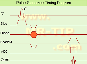
(GRE - sequence) A gradient echo is generated by using a pair of bipolar gradient pulses. In the pulse sequence timing diagram, the basic gradient echo sequence is illustrated. There is no refocusing 180° pulse and the data are sampled during a gradient echo, which is achieved by dephasing the spins with a negatively pulsed gradient before they are rephased by an opposite gradient with opposite polarity to generate the echo.
See also the Pulse Sequence Timing Diagram. There you will find a description of the components.
The excitation pulse is termed the alpha pulse α. It tilts the magnetization by a flip angle α, which is typically between 0° and 90°. With a small flip angle there is a reduction in the value of transverse magnetization that will affect subsequent RF pulses.
The flip angle can also be slowly increased during data acquisition (variable flip angle: tilt optimized nonsaturation excitation).
The data are not acquired in a steady state, where z-magnetization recovery and destruction by ad-pulses are balanced.
However, the z-magnetization is used up by tilting a little more of the remaining z-magnetization into the xy-plane for each acquired imaging line.
Gradient echo imaging is typically accomplished by examining the FID, whereas the read gradient is turned on for localization of the signal in the readout direction. T2* is the characteristic decay time constant associated with the FID. The contrast and signal generated by a gradient echo depend on the size of the longitudinal magnetization and the flip angle.
When α = 90° the sequence is identical to the so-called partial saturation or saturation recovery pulse sequence.
In standard GRE imaging, this basic pulse sequence is repeated as many times as image lines have to be acquired.
Additional gradients or radio frequency pulses are introduced with the aim to spoil to refocus the xy-magnetization at the moment when the spin system is subject to the next α pulse.
As a result of the short repetition time, the z-magnetization cannot fully recover and after a few initial α pulses there is an equilibrium established between z-magnetization recovery and z-magnetization reduction due to the α pulses.
Gradient echoes have a lower SAR, are more sensitive to field inhomogeneities and have a reduced crosstalk, so that a small or no slice gap can be used.
In or out of phase imaging depending on the selected TE (and field strength of the magnet) is possible.
As the flip angle is decreased, T1 weighting can be maintained by reducing the TR.
T2* weighting can be minimized by keeping the TE as short as possible, but pure T2 weighting is not possible.
By using a reduced flip angle, some of the magnetization value remains longitudinal (less time needed to achieve full recovery) and for a certain T1 and TR, there exist one flip angle that will give the most signal, known as the "Ernst angle".
Contrast values:
PD weighted: Small flip angle (no T1), long TR (no T1) and short TE (no T2*)
T1 weighted: Large flip angle (70°), short TR (less than 50ms) and short TE
T2* weighted: Small flip angle, some longer TR (100 ms) and long TE (20 ms)
Classification of GRE sequences can be made into four categories:
See also Gradient Recalled Echo Sequence, Spoiled Gradient Echo Sequence, Refocused Gradient Echo Sequence, Ultrafast Gradient Echo Sequence.
| | | | | |
• View the DATABASE results for 'Gradient Echo Sequence' (70).
| | | | | | Further Reading: | Basics:
|
|
News & More:
| |
| |
| | | Searchterm 'Contrast Enhanced' was also found in the following services: | | | | |
| | |
| |
|

From Philips Medical Systems;
the Intera-family offers with this member a wide range of possibilities, efficiency and a ergonomic and intuitive serving-platform. Also available as Intera CV for cardiac and Intera I/T for interventional MR procedures.
The scanners are also equipped with SENSE technology, which is essential for high-quality contrast enhanced magnetic resonance angiography, interactive cardiac MR and diffusion tensor imaging ( DTI) fiber tracking.
The increased accuracy and clarity of MR scans obtained with this technology allow for faster and more accurate diagnosis of potential problems like patient friendliness and expands the breadth of applications including cardiology, oncology and interventional MR.
Device Information and Specification
CLINICAL APPLICATION
Whole body
CONFIGURATION
Short bore compact
Standard: head, body, C1, C3; Optional: Small joint, flex-E, flex-R, endocavitary (L and S), dual TMJ, knee, neck, T/L spine, breast; Optional phased array: Spine, pediatric, 3rd party connector; Optional SENSE coils: Flex-S-M-L, flex body, flex cardiac
SE, Modified-SE ( TSE), IR (T1, T2, PD), STIR, FLAIR, SPIR, FFE, T1-FFE, T2-FFE, Balanced FFE, TFE, Balanced TFE, Dynamic, Keyhole, 3D, Multi Chunk 3D, Multi Stack 3D, K Space Shutter, MTC, TSE, Dual IR, DRIVE, EPI, Cine, 2DMSS, DAVE, Mixed Mode; Angiography: PCA, MCA, Inflow MRA, CE
TR
2.9 (Omni), 1.6 (Power), 1.6 (Master/Expl) msec
TE
1.0 (Omni), 0.7 (Power), 0.5 (Master/Expl) msec
RapidView Recon. greater than 500 @ 256 Matrix
0.1 mm(Omni), 0.05 mm (Pwr/Mstr/Expl)
128 x 128, 256 x 256,512 x 512,1024 x 1024 (64 for BOLD img.)
Variable in 1% increments
Lum.: 120 cd/m2; contrast: 150:1
Variable (op. param. depend.)
POWER REQUIREMENTS
380/400 V
| | | |
• View the DATABASE results for 'Intera 1.5T™' (2).
| | | | |
| | | | | |
| |
|
Liver imaging can be performed with sonography, computed tomography (CT) and magnetic resonance imaging ( MRI). Ultrasound is, caused by the easy access, still the first-line imaging method of choice; CT and MRI are applied whenever ultrasound imaging yields vague results. Indications are the characterization of metastases and primary liver tumors e.g., benign lesions such as focal nodular hyperplasia (FNH), adenoma, hemangioma and malignant lesions (cancer) such as hepatocellular carcinomas (HCC).
The decision, which medical imaging modality is more suitable, MRI or CT, is dependent on the different factors. CT is less costly and more widely available; modern multislice scanners provide high spatial resolution and short scan times but has the disadvantage of radiation exposure.
With the introduction of high performance MR systems and advanced sequences the image quality of MRI for the liver has gained substantially. Fast spin echo or single shot techniques, often combined with fat suppression, are the most common T2 weighted sequences used in liver MRI procedures.
Spoiled gradient echo sequences are used as ideal T1 weighted sequences for evaluating of the liver. The repetition time (TR) can be sufficiently long to acquire enough sections covering the entire liver in one pass, and to provide good signal to noise. The TE should be the shortest in phase echo time (TE), which provides strong T1 weighting, minimizes magnetic susceptibility effects, and permits acquisition within one breath hold to cover the whole liver. A flip angle of 80° provides good T1 weighting and less of power deposition and tissue saturation than a larger flip angle that would provide comparable T1 weighting.
Liver MRI is very dependent on the administration of contrast agents, especially when detection and characterization of focal lesions are the issues. Liver MRI combined with MRCP is useful to evaluate patients with hepatic and biliary disease.
Gadolinium chelates are typical non-specific extracellular agents diffusing rapidly to the extravascular space of tissues being cleared by glomerular filtration at the kidney. These characteristics are somewhat problematic when a large organ with a huge interstitial space like the liver is imaged. These agents provide a small temporal imaging window (seconds), after which they begin to diffuse to the interstitial space not only of healthy liver cells but also of lesions, reducing the contrast gradient necessary for easy lesion detection. Dynamic MRI with multiple phases after i.v. contrast media (Gd chelates), with arterial, portal and late phase images (similar to CT) provides additional information.
An additional advantage of MRI is the availability of liver-specific contrast agents (see also Hepatobiliary Contrast Agents). Gd-EOB-DTPA (gadoxetate disodium, Gadolinium ethoxybenzyl dimeglumine, EOVIST Injection, brand name in other countries is Primovist) is a gadolinium-based MRI contrast agent approved by the FDA for the detection and characterization of known or suspected focal liver lesions.
Gd-EOB-DTPA provides dynamic phases after intravenous injection, similarly to non-specific gadolinium chelates, and distributes into the hepatocytes and bile ducts during the hepatobiliary phase. It has up to 50% hepatobiliary excretion in the normal liver.
Since ferumoxides are not eliminated by the kidney, they possess long plasmatic half-lives, allowing circulation for several minutes in the vascular space. The uptake process is dependent on the total size of the particle being quicker for larger particles with a size of the range of 150 nm (called superparamagnetic iron oxide). The smaller ones, possessing a total particle size in the order of 30 nm, are called ultrasmall superparamagnetic iron oxide particles and they suffer a slower uptake by RES cells. Intracellular contrast agents used in liver MRI are primarily targeted to the normal liver parenchyma and not to pathological cells. Currently, iron oxide based MRI contrast agents are not marketed.
Beyond contrast enhanced MRI, the detection of fatty liver disease and iron overload has clinical significance due to the potential for evolution into cirrhosis and hepatocellular carcinoma. Imaging-based liver fat quantification (see also Dixon) provides noninvasively information about fat metabolism; chemical shift imaging or T2*-weighted imaging allow the quantification of hepatic iron concentration.
See also Abdominal Imaging, Primovistâ„¢, Liver Acquisition with Volume Acquisition (LAVA), T1W High Resolution Isotropic Volume Examination (THRIVE) and Bolus Injection.
For Ultrasound Imaging (USI) see Liver Sonography at Medical-Ultrasound-Imaging.com. | | | | | | | | | | |
• View the DATABASE results for 'Liver Imaging' (13).
| | |
• View the NEWS results for 'Liver Imaging' (10).
| | | | | | Further Reading: | Basics:
|
|
News & More:
|  |
Utility and impact of magnetic resonance elastography in the clinical course and management of chronic liver disease
Saturday, 20 January 2024 by www.nature.com | | |
Even early forms of liver disease affect heart health, Cedars-Sinai study finds
Thursday, 8 December 2022 by www.eurekalert.org | | |
For monitoring purposes, AI-aided MRI does what liver biopsy does with less risk, lower cost
Wednesday, 28 September 2022 by radiologybusiness.com | | |
Perspectum: High Liver Fat (Hepatic Steatosis) Linked to Increased Risk of Hospitalization in COVID-19 Patients With Obesity
Monday, 29 March 2021 by www.businesswire.com | | |
EMA's final opinion confirms restrictions on use of linear gadolinium agents in body scans
Friday, 21 July 2017 by www.ema.europa.eu | | |
T2-Weighted Liver MRI Using the MultiVane Technique at 3T: Comparison with Conventional T2-Weighted MRI
Friday, 16 October 2015 by www.ncbi.nlm.nih.gov | | |
EORTC study aims to qualify ADC as predictive imaging biomarker in preoperative regimens
Monday, 4 January 2016 by www.eurekalert.org | | |
MRI effectively measures hemochromatosis iron burden
Saturday, 3 October 2015 by medicalxpress.com | | |
Total body iron balance: Liver MRI better than biopsy
Sunday, 15 March 2015 by www.eurekalert.org |
|
| |
| | | | |
| | | |
|
| |
 | Look
Ups |
| |


