 | Info
Sheets |
| | | | | | | | | | | | | | | | | | | | | | | | |
 | Out-
side |
| | | | |
|
| | | | | |  | Searchterm 'Imaging' was also found in the following services: | | | | |
|  |  |
| |
|

Founded in 1991, ImaRx Pharmaceutical Corp. designs, develops and markets pharmaceuticals for medical imaging ( MRI, ultrasound and computed tomography) for the radiological imaging industry.
ImaRx Pharmaceutical Corp., announced 1999 that it has been acquired by E.I DuPont de Nemours and Co., Inc.. The terms of the acquisition provide a royalty-free licensing arrangement with a newly-formed company, ImaRx LLC ("LLC"), to pursue and develop new products and technologies for drug and gene delivery independent from DuPont.
Yamanouchi Pharmaceutical Co. Ltd., ImaRx' licensee for Asian territories for this product, will continue to develop the product in Asia as DuPont's licensee. ImaRx LLC will have ownership of all other targeted and therapeutic products previously owned by ImaRx, including imaging products outside of diagnostic ultrasound imaging and two other imaging products, SonoRx® and LumenHance®, which are both FDA approved and licensed to BRACCO Diagnostics.
MRI Contrast Agents:
Contact Information
MAIL
ImaRx LLC
1635 East 18th Street
Tucson AZ 85719-6803
USA
| | | | | |
| | | | | |
| |
|
Knee MRI, with its high soft tissue contrast is one of the main imaging tools to depict knee joint pathology. MRI allows accurate imaging of intra-articular structures such as ligaments, cartilage, menisci, bone marrow, synovium, and adjacent soft tissue.
Knee exams require a dedicated extremity coil, providing a homogenous imaging volume and high SNR to ensure best signal coverage.
A complete knee MR examination includes for example sagittal and coronal T1 weighted, and proton density weighted pulse sequences +/- fat saturation, or STIR sequences. For high spatial resolution, maximal 4 mm thick slices with at least an in plane resolution of 0.75 mm and small gap are recommended. To depict the anterior cruciate ligament clearly, the sagittal plane has to be rotated 10 - 20° externally (parallel to the medial border of the femoral condyle). Retropatellar cartilage can bee seen for example in axial T2 weighted gradient echo sequences with Fatsat. However, the choice of the pulse sequences is depended of the diagnostic question, the used scanner, and preference of the operator.
Diagnostic quality in knee imaging is possible with field strengths ranging from 0.2 to 3T. With low field strengths more signal averages must be measured, resulting in increased scan times to provide equivalent quality as high field strengths.
More diagnostic information of meniscal tears and chondral defects can be obtained by direct magnetic resonance arthrography, which is done by introducing a dilute solution of gadolinium in saline (1:1000) into the joint capsule. The knee is then scanned in all three planes using T1W sequences with fat suppression. For indirect arthrography, the contrast is given i.v. and similar scans are started 20 min. after injection and exercise of the knee.
Frequent indications of MRI scans in musculoskeletal knee diseases are: e.g., meniscal degeneration and tears, ligament injuries, osteochondral fractures, osteochondritis dissecans, avascular bone necrosis and rheumatoid arthritis. See also Imaging of the Extremities and STIR. | | | | | |
• View the DATABASE results for 'Knee MRI' (4).
| | |
• View the NEWS results for 'Knee MRI' (4).
| | | | |  Further Reading: Further Reading: | | Basics:
|
|
News & More:
| |
| |
| | | | | |
| |
|
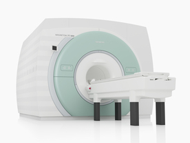
From Siemens Medical Systems;
The MAGNETOM 7T is designed as an open research platform. 7T MRI provides anatomical detail at the submillimiter scale, enhanced contrast mechanisms, outstanding spectroscopy performance, ultra-high resolution functional imaging ( fMRI), multinuclear whole-body MRI and functional information.
This ultra high field (UHF) MRI device is a research system and not cleared, approved or licensed in any jurisdiction for patient examinations.
Device Information and Specification
CLINICAL APPLICATION
Whole body
High-performance, ultra high field coils available. Integration and support for coil developments.
CHANNELS (min. / max. configuration)
32, optional 8 channels TX array
40 x 40 x 30 cm³ less than 8% nonlinearity
MAGNET WEIGHT (gantry included)
35017 kg
DIMENSION H*W*D (gantry included)
320 x 240 x 317,5 cm
MAX. AMPLITUDE
up to 70 mT/m
Up to 3rd order shim coils, user configurable B0 shim ? B0 maps and ROI definition
POWER REQUIREMENTS
2000 Volts, 650A
| | | | | | | Further Reading: | | Basics:
|
|
News & More:
| |
| |
| | | Searchterm 'Imaging' was also found in the following services: | | | | |
| | |
| |
|
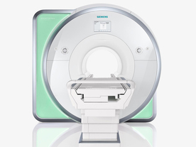
From Siemens Medical Systems;
Received FDA clearance in 2010.
The MAGNETOM Aera is a patient friendly, comfortable 1.5 Tesla MRI system with advanced radio frequency chain.
The system is equipped with the Tim 4G and Dot system (Total imaging matrix + Day optimizing throughput), to enhance both productivity and image quality.
Tim 4G technology provides improved SNR. The standard system configuration of 48 radio frequency channels and 204 coil elements creates an imaging matrix that allows maximum use of coil elements at full field of view. Dot provides improved image consistency through new features like auto align, auto FoV and automatic bolus detection.
Device Information and Specification
CLINICAL APPLICATION
Whole body
Head, spine, torso/ body coil, neurovascular, cardiac, neck, shoulder, knee, wrist, foot//ankle and multi-purpose flex coils. Peripheral vascular, breast, shoulder. Up to 60% more SNR with Tim 4G.
CHANNELS (min. / max. configuration)
48, 64
MINIMUM TE
3-D GRE: 0.22 (256 matrix), Ultra-short TE
At isocenter: L-R 70 cm, A-P (with table) 55 cm
MAGNET WEIGHT (gantry included)
3121 kg
DIMENSION H*W*D (gantry included)
145 x 231 x 219 cm
MAX. AMPLITUDE
33 or 45 mT/m
3 linear with 20 coils, 5 nonlinear 2nd-order
POWER REQUIREMENTS
380 / 400 / 420 / 440 / 460 / 480 V, 3-phase + ground; 85 kVA
| | | | | |
| | | | | |
| |
|
The MRI device is located within a specially shielded room ( Faraday cage) to avoid outside interference, caused by the use of radio waves very close in frequency to those of ordinary FM radio stations.
The MRI procedure can easily be performed through clothing and bones, but attention must be paid to ferromagnetic items, because they will be attracted from the magnetic field. A hospital gown is appropriate, or the patient should wear clothing without metal fasteners and remove any metallic objects like hairpins, jewelry, eyeglasses, clocks, hearing aids, any removable dental work, lighters, coins etc., not only for MRI safety reasons.
Metal in or around the scanned area can also cause errors in the reconstructed images ( artifacts). Because the strong magnetic field can displace, or disrupt metallic objects, people with an implanted active device like a cardiac pacemaker cannot be scanned under normal circumstances and should not enter the MRI area.
The MRI machine can look like a short tunnel or has an open MRI design and the magnet does not completely surround the patient. Usually the patient lies on a comfortable motorized table, which slides into the scanner, depending on the MRI device, patients may be also able to sit up. If a contrast agent is to be administered, intravenous access will be placed. A technologist will operate the MRI machine and observe the patient during the examination from an adjacent room. Several sets of images are usually required, each taking some minutes. A typical MRI scan includes three to nine imaging sequences and may take up to one hour. Improved MRI devices with powerful magnets, newer software, and advanced sequences may complete the process in less time and better image quality.
Before and after the most MRI procedures no special preparation, diet, reduced activity, and extra medication is necessary. The magnetic field and radio waves are not felt and no pain is to expect.
Movement can blur MRI images and cause certain artifacts. A possible problem is the claustrophobia that some patients experience from being inside a tunnel-like scanner. If someone is very anxious or has difficulty to lie still, a sedative agent may be given. Earplugs and/or headphones are usually given to the patient to reduce the loud acoustic noise, which the machine produces during normal operation. A technologist observes the patient during the test. Some MRI scanners are equipped with televisions and music to help the examination time pass.
MRI is not a cheap examination, however cost effective by eliminating the need for invasive radiographic procedures, biopsies, and exploratory surgery. MRI scans can also save money while minimizing patient risk and discomfort. For example, MRI can reduce the need for X-ray angiography and myelography, and can eliminate unnecessary diagnostic procedures that miss occult disease. See also Magnetic Resonance Imaging MRI, Medical Imaging, Cervical Spine MRI, Claustrophobia, MRI Risks and Pregnancy.
For Ultrasound Imaging (USI) see Ultrasound Imaging Procedures at Medical-Ultrasound-Imaging.com.
See also the related poll result: ' MRI will have replaced 50% of x-ray exams by' | | | | | |
• View the DATABASE results for 'MRI Procedure' (11).
| | |
• View the NEWS results for 'MRI Procedure' (6).
| | | | | | Further Reading: | News & More:
|
|
| |
| | | | |
| | | |
|
| |
 | Look
Ups |
| |


