 | Info
Sheets |
| | | | | | | | | | | | | | | | | | | | | | | | |
 | Out-
side |
| | | | |
|
| | | | | |  | Searchterm 'cardiac' was also found in the following services: | | | | |
|  |  |
| |
|

It is important to remember when working around a superconducting magnet that the magnetic field is always on. Under usual working conditions the field is never turned off. Attention must be paid to keep all ferromagnetic items at an adequate distance from the magnet. Ferromagnetic objects which came accidentally under the influence of these strong magnets can injure or kill individuals in or nearby the magnet, or can seriously damage every hardware, the magnet itself, the cooling system, etc..
See MRI resources Accidents.
The doors leading to a magnet room should be closed at all times except when entering or exiting the room. Every person working in or entering the magnet room or adjacent rooms with a magnetic field has to be instructed about the dangers. This should include the patient, intensive-care staff, and maintenance-, service- and cleaning personnel, etc..
The 5 Gauss limit defines the 'safe' level of static magnetic field exposure. The value of the absorbed dose is fixed by the authorities to avoid heating of the patient's tissue and is defined by the specific absorption rate.
Leads or wires that are used in the magnet bore during imaging procedures, should not form large-radius wire loops. Leg-to-leg and leg-to-arm skin contact should be prevented in order to avoid the risk of burning due to the generation of high current loops if the legs or arms are allowed to touch. The patient's skin should not be in contact with the inner bore of the magnet.
The outflow from cryogens like liquid helium is improbable during normal operation and not a real danger for patients.
The safety of MRI contrast agents is tested in drug trials and they have a high compatibility with very few side effects. The variations of the side effects and possible contraindications are similar to X-ray contrast medium, but very rare. In general, an adverse reaction increases with the quantity of the MRI contrast medium and also with the osmolarity of the compound.
See also 5 Gauss Fringe Field, 5 Gauss Line, Cardiac Risks, Cardiac Stent, dB/dt, Legal Requirements, Low Field MRI, Magnetohydrodynamic Effect, MR Compatibility, MR Guided Interventions, Claustrophobia, MRI Risks and Shielding. | | | | | | | | | • For this and other aspects of MRI safety see our InfoSheet about MRI Safety. | | | • Patient-related information is collected in our MRI Patient Information.
| | | | | | | | | |  Further Reading: Further Reading: | | Basics:
|
|
News & More:
| |
| |
|  |  | MRI Safety Resources | | | | |
|  |  |  |
| |
|
(LE) Myocardial late enhancement in contrast enhanced cardiac MRI has the ability to precisely delineate myocardial scar associated with coronary artery disease. Viability imaging implies evaluating infarcted myocardium to see whether there is enough viable tissue available for revascularization. The reversal of myocardial dysfunction is particularly relevant in patients with depressed ventricular function because revascularization improves long-term survival. In comparison to SPECT and PET imaging, myocardial late enhancement MRI demonstrates areas of delayed enhancement exactly in correlation with the infarcted region.
Viability on cardiac MRI (CMR) is based on the fact that all infarcts enhance vividly 10-15 minutes after the administration of intravenous paramagnetic contrast agents. This enhancement represents the accumulation of gadolinium in the extracellular space, due to the loss of membrane integrity in the infarcted tissue. This phenomenon of delayed hyperenhancement has been proven to correlate with the actual extent of the infarct.
MRI myocardial late enhancement can quantify the size, location and transmural extent of the infarct. If the transmural extent of the infarct (region of enhancement on MRI) is less than 50% of the wall thickness, there will be improved contractility in that segment following revascularization. In areas of hypokinesia, if there is a rim of "black" or non-infarcted myocardium that is not contracting well, it indicates the presence of hibernating myocardium, which is likely to improve after revascularization of the artery supplying that particular territory.
The total duration of a myocardial late enhancement MR imaging protocol for viability is approximately 30 minutes, including scout images, first-pass images, cine images in two planes, and delayed myocardial enhancement images. In order to assess viable myocardium, the gadolinium contrast agent is injected at a dose of 0.15 to 0.2 mmol/kg. After about 10 minutes, short axis and long axis views (see cardiac axes) of the heart are obtained using an inversion prepared ECG gated gradient echo sequence. The inversion pulse is adjusted to suppress normal myocardium. Areas of nonviable myocardium retain extremely high signal intensity, black areas show normal tissue.
For Ultrasound Imaging (USI) see Myocardial Contrast Echocardiography at Medical-Ultrasound-Imaging.com. | |  | |
• View the DATABASE results for 'Myocardial Late Enhancement' (6).
| | | | |  Further Reading: Further Reading: | Basics:
|
|
News & More:
| |
| |
|  | |  |  |  |
| |
|

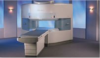
From Hitachi Medical Systems America, Inc.;
the AIRIS made its debut in 1995. Hitachi followed up with the AIRIS II system, which has proven equally successfully. 'All told, Hitachi has installed more than 1,000 MRI systems in the U.S., holding more than 17 percent of the total U.S. MRI installed base, and more than half of the installed base of open MR systems,' says Antonio Garcia, Frost and Sullivan industry research analyst.
Now Altaire employs a blend of innovative Hitachi features called VOSI™ technology, optimizing each sub-system's performance in concert with the
other sub-systems, to give the seamless mix of high-field performance
and the patient comfort, especially for claustrophobic patients, of open MR systems.
Device Information and Specification
CLINICAL APPLICATION
Whole body
DualQuad T/R Body Coil, MA Head, MA C-Spine, MA Shoulder, MA Wrist, MA CTL Spine, MA Knee, MA TMJ, MA Flex Body (3 sizes), Neck, small and large Extremity, PVA (WIP), Breast (WIP), Neurovascular (WIP), Cardiac (WIP) and MA Foot//Ankle (WIP)
SE, GE, GR, IR, FIR, STIR, ss-FSE, FSE, DE-FSE/FIR, FLAIR, ss/ms-EPI, ss/ms EPI- DWI, SSP, MTC, SE/GE-EPI, MRCP, SARGE, RSSG, TRSG, BASG, Angiography: CE, PC, 2D/3D TOF
IMAGING MODES
Single, multislice, volume study
TR
SE: 30 - 10,000msec GE: 3.6 - 10,000msec IR: 50 - 16,700msec FSE: 200 - 16,7000msec
TE
SE : 8 - 250msec IR: 5.2 -7,680msec GE: 1.8 - 2,000 msec FSE: 5.2 - 7,680
0.05 sec/image (256 x 256)
2D: 2 - 100 mm; 3D: 0.5 - 5 mm
Level Range: -2,000 to +4,000
COOLING SYSTEM TYPE
Water-cooled
3.1 m lateral, 3.6 m vertical
| |  | |
• View the DATABASE results for 'Altaire™' (2).
| | | | |  Further Reading: Further Reading: | News & More:
|
|
| |
|  |  | Searchterm 'cardiac' was also found in the following services: | | | | |
|  |  |
| |
|
With this method irregular RR intervals in cardiac gating during cardiovascular imaging are rejected and then repeated to improve the image quality, whereby the cardiac frequency is used as a basis of the normal heart rate.
The RR interval window determines the percentage variation of the heart rate. Variations of the acquired data outside the window are rejected and not used in the image reconstruction. Also one interval after the arrhytmic beat will be rejected.
Arrhythmia rejection may be inappropriate for patients with certain pathologies, because if the RR interval is constant long, short, long, - all intervals would be rejected. Also a disadvantage is the time consume, but in some cases this function is mandatory, e.g. for diverse retrospective triggered sequences. | |  | | | |  Further Reading: Further Reading: | Basics:
|
|
News & More:
| |
| |
|  | |  |  |  |
| |
|
| | | |  | |
• View the DATABASE results for 'Black Blood MRA' (6).
| | | | |
|  | |  |  |
|  | | |
|
| |
 | Look
Ups |
| |