Magnetic Resonance - Technology Information Portal Welcome to MRI Technology
Info
  Sheets

Out-
      side


 'Breath Hold Imaging' 
SEARCH FOR    
 
  2 3 5 A B C D E F G H I J K L M N O P Q R S T U V W X Y Z
Result : Searchterm 'Breath Hold Imaging' found in 1 term [] and 6 definitions [], (+ 14 Boolean[] results
previous     6 - 10 (of 21)     next
Result Pages : [1]  [2]  [3 4 5]
MRI Resources 
Directories - Databases - General - Colonography - Jobs - Health
 
In Phase Image
 
The term in phase refers to an image in which the signals from two spectral components (such as fat and water) add constructively in a voxel. T1 weighted in phase images are acquired by a gradient echo-based technique with a short TR, TE and a high flip angle greater than 60 degrees. To some degree, in phase sequences are more sensitive to detection of focal hepatic lesions than out of phase for evaluating reduced lesion-to-liver contrast, but the choice for a T1 gradient echo sequence is still based on field strength, advanced imaging techniques (breath hold imaging), and physician preference.
 
Images, Movies, Sliders:
 MRI Liver In Phase  Open this link in a new window
    
 
spacer
 
Further Reading:
  News & More:
Direct Water and Fat Determination in Two-Point Dixon Imaging
April 2013   by scholarship.rice.edu    
MRI Resources 
Contrast Enhanced MRI - MRCP - Homepages - Absorption and Emission - Open Directory Project - Anatomy
 
Volumetric Imaging
 
Volumetric imaging is a 3D technique where all the MRI signals are collected from the entire tissue sample and imaged as a whole entity, therefore providing a high signal to noise ratio. The acquisition of isotropic voxels or thin slices with high spatial resolution allows to create multiplanar reconstructions in all planes; a compensation for the usually longer scan time. The acquisition time can be reduced by parallel imaging technique.
New T2 weighted variants of 3D sequences (FSE-XETA, T2-SPACE, VISTA) have been introduced that differ from conventional FSE sequences. An echo train containing up to 200 echoes obtained at a minimum echo spacing allows very fast acquisition. A flip angle modulation (flip angle sweep - FAS) during the FSE readout carries magnetization as long as possible to avoid blurring and provide optimal signal at the effective echo time. This type of imaging is well suited for brain and spine MRI procedures.
Newer T1 weighted variants include Liver Acquisition with Volume Acquisition (LAVA) and T1W High Resolution Isotropic Volume Examination (THRIVE), which have advantages for dynamic breath hold imaging in liver and abdominal examinations.

See also Volume Imaging, 3 Dimensional Imaging.
 
Images, Movies, Sliders:
 Brain MRI Sagittal T1 001  Open this link in a new window
    
 Circle of Willis, Time of Flight, MIP  Open this link in a new window
    
SlidersSliders Overview

 MRI of the Skull Base  Open this link in a new window
    
SlidersSliders Overview

 
spacer

• View the DATABASE results for 'Volumetric Imaging' (4).Open this link in a new window


• View the NEWS results for 'Volumetric Imaging' (1).Open this link in a new window.
 
Further Reading:
  Basics:
Cutting Edge Imaging of THE Spine
February 2007   by www.pubmedcentral.nih.gov    
3-D VOLUMETRIC IMAGING FOR STEREOTACTIC LESIONAL AND DEEP BRAIN STIMULATION SURGERY
MRI Resources 
Developers - PACS - MRI Accidents - Contrast Enhanced MRI - MRCP - Spectroscopy pool
 
Signa HDx 3.0Tâ„¢InfoSheet: - Devices -
Intro, 
Types of Magnets, 
Overview, 
etc.
 
gehealthcare.com/euen/mri/products/signa-hdx-3t/index.html From GE Healthcare;
The Signa HDx MRI system is GE's leading edge whole body magnetic resonance scanner designed to support high resolution, high signal to noise ratio, and short scan times.
Signa HDx 3.0T offers new technologies like ultra-fast image reconstruction through the new XVRE recon engine, advancements in parallel imaging algorithms and the broadest range of premium applications. The HD applications, PROPELLER (high-quality brain imaging extremely resistant to motion artifacts), TRICKS (contrast-enhanced angiographic vascular lower leg imaging), VIBRANT (for breast MRI), LAVA (high resolution liver imaging with shorter breath holds and better organ coverage) and MR Echo (high-definition cardiac images in real time) offer unique capabilities.
Device Information and Specification
CLINICAL APPLICATION
Whole body
CONFIGURATION
Compact short bore
Head and body coil, T/R quadrature head; optional coils e.g., T/R phased array extremity abdomen, spine, breast, knee, shoulder, cardiac imaging coils
SYNCHRONIZATION
ECG/peripheral, respiratory gating
PULSE SEQUENCES
SE, IR, 2D/3D GRE, RF-spoiled GRE, 2DFGRE, 2DFSPGR, 3DFGRE, 3DFSPGR, 3DTOFGRE, 3DFSPGR, 2DFSE, 2DFSE-XL, 2DFSE-IR, T1-FLAIR, SSFSE, EPI, DW-EPI, BRAVO, Angiography: 2D/3D TOF, 2D/3D phase contrast vascular
IMAGING MODES
Single, multislice, volume study, fast scan, multi slab, cine, localizer
1 cm to 40 cm continuous
2D 0.5 mm; 3D 0.1 mm
1024 x 1024
PIXEL INTENSITY
256 gray levels
60 cm
MAGNET WEIGHT
12000 kg
H*W*D
240 x 2216,6 x 201,6 cm
POWER REQUIREMENTS
480 or 380/415, 3 phase ||
COOLING SYSTEM TYPE
Closed-loop water-cooled grad.
0.03 L/hr helium
STRENGTH
23 - 50 mT/m
80 - 150 mT/m/ms
5-GAUSS FRINGE FIELD
2.8 m / 5.0 m
second and high order
spacer
MRI Resources 
MRA - MR Myelography - Spectroscopy - Devices - Jobs - Research Labs
 
MAGNETOM Aera
 
www.healthcare.siemens.com/magnetic-resonance-imaging/0-35-to-1-5t-mri-scanner/magnetom-aera/ 
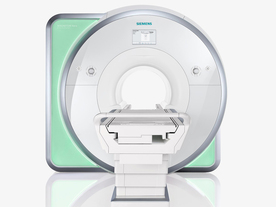
From Siemens Medical Systems;
Received FDA clearance in 2010.
The MAGNETOM Aera is a patient friendly, comfortable 1.5 Tesla MRI system with advanced radio frequency chain.
The system is equipped with the Tim 4G and Dot system (Total imaging matrix + Day optimizing throughput), to enhance both productivity and image quality.
Tim 4G technology provides improved SNR. The standard system configuration of 48 radio frequency channels and 204 coil elements creates an imaging matrix that allows maximum use of coil elements at full field of view. Dot provides improved image consistency through new features like auto align, auto FoV and automatic bolus detection.
Device Information and Specification
CLINICAL APPLICATION
Whole body
CONFIGURATION
Open bore
Head, spine, torso/ body coil, neurovascular, cardiac, neck, shoulder, knee, wrist, foot//ankle and multi-purpose flex coils. Peripheral vascular, breast, shoulder. Up to 60% more SNR with Tim 4G.
CHANNELS (min. / max. configuration)
48, 64
IMAGING TECHNIQUES
iPAT, mSENSE and GRAPPA (image, k-space), noncontrast angiography, plaque imaging, radial motion compensation, Dixon, improved workflow with Dot, Caipirinha - single digit breath-holds for 3-D body imaging.
MINIMUM TR
3-D GRE: 0.95 (256 matrix)
MINIMUM TE
3-D GRE: 0.22 (256 matrix), Ultra-short TE
FOV
0.5 - 50
BORE DIAMETER
or W x H
At isocenter: L-R 70 cm, A-P (with table) 55 cm
TABLE CAPACITY
250 kg
MAGNET WEIGHT (gantry included)
3121 kg
DIMENSION H*W*D (gantry included)
145 x 231 x 219 cm
5-GAUSS FRINGE FIELD
2.5 m / 4.0 m
CRYOGEN USE
Zero boil off rate, approx. 10 years
COOLING SYSTEM
Water
up to 200 T/m/s
MAX. AMPLITUDE
33 or 45 mT/m
3 linear with 20 coils, 5 nonlinear 2nd-order
POWER REQUIREMENTS
380 / 400 / 420 / 440 / 460 / 480 V, 3-phase + ground; 85 kVA
spacer
MRI Resources 
Journals - Spine MRI - Bioinformatics - Health - Resources - MRI Technician and Technologist Schools
 
Lung ImagingMRI Resource Directory:
 - Lung Imaging -
 
Lung imaging is furthermore a challenge in MRI because of the predominance of air within the lungs and associated susceptibility issues as well as low signal to noise of the inflated lung parenchyma. Cardiac and respiratory triggered or breath hold sequences allow diagnostic imaging, however a comparable image quality with computed tomography is still difficult to achieve.
Assumptions for lung MRI:
•
Low signal to noise ratio of the inherently low lung proton density.
•
Cardiac and respiratory motion artifacts.
•
Magnetic susceptibility effects of large magnetic field gradients.
•
Very short transverse relaxation times and significant diffusion yielding short T2 (30-70 msec), short T2* (1-3 msec), and additional long T1 relaxation times (1300-1500 msec).
•
The extreme short T2 values are responsible for a fast signal decay during a single shot readout, resulting in blurring.

The current trends in MRI are the use of new imaging technologies and increasingly powerful magnetic fields. Among these technologies are parallel imaging techniques as well as ventilation agents like hyperpolarized helium for the use as an inert inhalational contrast agent to study lung ventilation properties. With hyperpolarized gases clear images of the lungs can be obtained without using a large magnetic field (see also back projection imaging). Single shot sequences (e.g. TSE or Half Fourier Acquisition Single Shot Turbo Spin Echo HASTE) used in lung MR imaging benefits from parallel imaging techniques due to reduced relaxation time effects during the echo train and therefore reduced image blurring as well as reduced motion artifacts.
In the future, more effective contrast agents may provide an alternative solution to the need for high field MRI. Dynamic contrast enhanced MRI perfusion has demonstrated a potential in the diagnosis of pulmonary embolism or to characterize lung cancer and mediastinal tumors. 3D contrast enhanced magnetic resonance angiography of the thoracic vessel.

See also the related poll result: 'MRI will have replaced 50% of x-ray exams by'
 
Images, Movies, Sliders:
 Anatomic Imaging of the Lungs  Open this link in a new window
      

Courtesy of  Robert R. Edelman
 Normal Lung Gd Perfusion MRI  Open this link in a new window
      

Courtesy of  Robert R. Edelman

 MRI Thorax Basal Plane  Open this link in a new window
 
Radiology-tip.comradLung Scintigraphy
spacer

• View the DATABASE results for 'Lung Imaging' (7).Open this link in a new window


• View the NEWS results for 'Lung Imaging' (3).Open this link in a new window.
 
Further Reading:
  Basics:
A safer approach for diagnostic medical imaging
Monday, 29 September 2014   by www.eurekalert.org    
Parallel Lung Imaging(.pdf)
  News & More:
Chest MRI a viable alternative to chest CT in COVID-19 pneumonia follow-up
Monday, 21 September 2020   by www.healthimaging.com    
CT Imaging Features of 2019 Novel Corona virus (2019-nCoV)
Tuesday, 4 February 2020   by pubs.rsna.org    
Polarean Imaging Phase III Trial Results Point to Potential Improvements in Lung Imaging
Wednesday, 29 January 2020   by www.diagnosticimaging.com    
Low Power MRI Helps Image Lungs, Brings Costs Down
Thursday, 10 October 2019   by www.medgadget.com    
Chest MRI Using Multivane-XD, a Novel T2-Weighted Free Breathing MR Sequence
Thursday, 11 July 2019   by www.sciencedirect.co    
Researchers Review Importance of Non-Invasive Imaging in Diagnosis and Management of PAH
Wednesday, 11 March 2015   by lungdiseasenews.com    
New MRI Approach Reveals Bronchiectasis' Key Features Within the Lung
Thursday, 13 November 2014   by lungdiseasenews.com    
MRI techniques improve pulmonary embolism detection
Monday, 19 March 2012   by medicalxpress.com    
  News & More:
Partnership with VIDA to streamline adoption of advanced MRI of the lungs
Monday, 11 September 2023   by www.itnonline.com    
MRI Resources 
MRCP - Brain MRI - Spine MRI - Safety Products - Education - NMR
 
previous      6 - 10 (of 21)     next
Result Pages : [1]  [2]  [3 4 5]
 Random Page
 
Share This Page
FacebookTwitterLinkedIn

MR-TIP    
Community   
User
Pass
Forgot your UserID/Password ?    


How AI will impact MRI :
only diagnostics 
saving time 
reducing cost 
makes planning obsolete 
reduce human knowledge 
not at all 

Look
      Ups


MR-TIP.com uses cookies! By browsing MR-TIP.com, you agree to our use of cookies.

Magnetic Resonance - Technology Information Portal
Member of SoftWays' Medical Imaging Group - MR-TIP • Radiology-TIP • Medical-Ultrasound-Imaging • 
Copyright © 2003 - 2024 SoftWays. All rights reserved. [ 6 May 2024]
Terms of Use | Privacy Policy | Advertising
 [last update: 2024-02-26 03:41:00]