Magnetic Resonance - Technology Information Portal Welcome to MRI Technology
Info
  Sheets

Out-
      side


 'Decay' 
SEARCH FOR    
 
  2 3 5 A B C D E F G H I J K L M N O P Q R S T U V W X Y Z
Result : Searchterm 'Decay' found in 3 terms [] and 22 definitions []
previous     6 - 10 (of 25)     next
Result Pages : [1]  [2 3 4 5]
Searchterm 'Decay' was also found in the following services: 
spacer
Forum  (3)  
 
Coherent Gradient EchoInfoSheet: - Sequences - 
Intro, 
Overview, 
Types of, 
etc.MRI Resource Directory:
 - Sequences -
 
Coherent gradient echo sequences can measure the free induction decay (FID), generated just after each excitation pulse or the echo formed prior to the next pulse. Coherent gradient echo sequences are very sensitive to magnetic field inhomogeneity. An alternative to spoiling is to incorporate residual transverse magnetization directly into the longitudinal steady state. These GRE sequences use a refocusing gradient in the phase encoding direction during the end module to maximize remaining transverse (xy) magnetization at the time when the next excitation is due, while the other two gradients are, in any case, balanced.
When the next excitation pulse is sent into the system with an opposed phase, it tilts the magnetization in the -a direction. As a result the z-magnetization is again partly tilted into the xy-plane, while the remaining xy-magnetization is tilted partly into the z-direction.
A fully refocused sequence with a properly selected and uniform f would yield higher signal, especially for tissues with long T2 relaxation times (high water content) so it is used in angiographic, myelographic or arthrographic examinations and is used for T2* weighting. The repetition time for this sequence has to be short. With short TR, coherent GE is also useable for breath hold and 3D technique. If the repetition time is about 200 msec there's no difference between spoiled or unspoiled GE. T1 weighting is better with spoiled techniques.
The common types include GRASS, FISP, FAST, and FFE.
The T2* component decreases with long TR and short TE. The T1 time is controlled by flip angle. The common TR is less than 50 ms and the common TE less than 15 ms
Other types have stronger T2 dependence but lower SNR. They include SSFP, CE-FAST, PSIF, and CE-FFE-T2.
Examples of fully refocused FID sequences are TrueFISP, bFFE and bTFE.
spacer
 
• Related Searches:
    • Spoiled Gradient Echo Sequence
    • Short Repetition Techniques
    • Incoherent Gradient Echo (Gradient Spoiled)
    • Transverse Magnetization
    • Dual Echo Sequence
Searchterm 'Decay' was also found in the following services: 
spacer
Radiology  (47) Open this link in a new windowUltrasound  (1) Open this link in a new window
Fast Relaxation Fast Spin EchoInfoSheet: - Sequences - 
Intro, 
Overview, 
Types of, 
etc.
 
(FRFSE, FR-FSE) The fast relaxation fast spin echo sequence provides high signal intensity of fluids even with short repetition times, and can be used with parallel imaging techniques for short breath hold imaging or respiratory gating for free-breathing, high isotropic resolution MR imaging. After signal decay at the end of the echo train, a negative 90° pulse align spins with long T2 from the transverse plane to the longitudinal plane, leading to a much faster recovery of tissues with long T2 time to the equilibrium and thus better contrast between tissues with long and short T2.
Fast relaxation FSE has advantages also for volumetric imaging as the TR can be substantially reduced and thus the scan time. The sequence can be post processed with maximum intensity projection, surface or volume rendering algorithms to visualize anatomical details in brain or spine MRI. Cerebro spinal fluid pulsation artifacts, often problematic in the cervical or thoracic spine may be reduced by radial sampling, in particular when combined with acquisitions of the PROPELLER type.

See also Fast spin echo, Driven Equilibrium.
 
Images, Movies, Sliders:
 Shoulder Sagittal T2 FatSat FRFSE  Open this link in a new window
    

Courtesy of  Robert R. Edelman
 Shoulder Axial T2 FatSat FRFSE  Open this link in a new window
 Shoulder Coronal T2 FatSat FRFSE  Open this link in a new window
    

Courtesy of  Robert R. Edelman
 
spacer
MRI Resources 
DICOM - Shoulder MRI - Mobile MRI Rental - Portals - Case Studies - Shielding
 
Fast Spin EchoForum -
related threadsInfoSheet: - Sequences - 
Intro, 
Overview, 
Types of, 
etc.
 
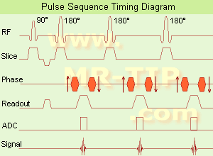
Fast Spin Echo Diagram (FSE) In the pulse sequence timing diagram, a fast spin echo sequence with an echo train length of 3 is illustrated. This sequence is characterized by a series of rapidly applied 180° rephasing pulses and multiple echoes, changing the phase encoding gradient for each echo.
The echo time TE may vary from echo to echo in the echo train. The echoes in the center of the K-space (in the case of linear k-space acquisition) mainly produce the type of image contrast, whereas the periphery of K-space determines the spatial resolution. For example, in the middle of K-space the late echoes of T2 weighted images are encoded. T1 or PD contrast is produced from the early echoes.
The benefit of this technique is that the scan duration with, e.g. a turbo spin echo turbo factor / echo train length of 9, is one ninth of the time. In T1 weighted and proton density weighted sequences, there is a limit to how large the ETL can be (e.g. a usual ETL for T1 weighted images is between 3 and 7). The use of large echo train lengths with short TE results in blurring and loss of contrast. For this reason, T2 weighted imaging profits most from this technique.
In T2 weighted FSE images, both water and fat are hyperintense. This is because the succession of 180° RF pulses reduces the spin spin interactions in fat and increases its T2 decay time. Fast spin echo (FSE) sequences have replaced conventional T2 weighted spin echo sequences for most clinical applications. Fast spin echo allows reduced acquisition times and enables T2 weighted breath hold imaging, e.g. for applications in the upper abdomen.
In case of the acquisition of 2 echoes this type of a sequence is named double fast spin echo / dual echo sequence, the first echo is usually density and the second echo is T2 weighted image. Fast spin echo images are more T2 weighted, which makes it difficult to obtain true proton density weighted images. For dual echo imaging with density weighting, the TR should be kept between 2000 - 2400 msec with a short ETL (e.g., 4).
Other terms for this technique are:
Turbo Spin Echo
Rapid Imaging Spin Echo,
Rapid Spin Echo,
Rapid Acquisition Spin Echo,
Rapid Acquisition with Refocused Echoes
 
Images, Movies, Sliders:
 Lumbar Spine T2 FSE Sagittal  Open this link in a new window
    

Courtesy of  Robert R. Edelman
 MRI - Anatomic Imaging of the Foot  Open this link in a new window
    
SlidersSliders Overview

 Lumbar Spine T2 FSE Axial  Open this link in a new window
    

Courtesy of  Robert R. Edelman
 
spacer

• View the DATABASE results for 'Fast Spin Echo' (31).Open this link in a new window

 
Further Reading:
  Basics:
MYELIN-SELECTIVE MRI: PULSE SEQUENCE DESIGN AND OPTIMIZATION
   by www.imaging.robarts.ca    
Advances in Magnetic Resonance Neuroimaging
Friday, 27 February 2009   by www.ncbi.nlm.nih.gov    
  News & More:
New MR sequence helps radiologists more accurately evaluate abnormalities of the uterus and ovaries
Thursday, 23 April 2009   by www.eurekalert.org    
Spin echoes, CPMG and T2 relaxation - Introductory NMR & MRI from Magritek
2013   by www.azom.com    
Searchterm 'Decay' was also found in the following services: 
spacer
Forum  (3)  
 
Fourier TransformationMRI Resource Directory:
 - Process Analysis -
 
(FT) The Fourier transformation is a mathematical procedure to separate out the frequency components of a signal from its amplitudes as a function of time, or the inverse Fourier transformation (IFT) calculates the time domain from the frequency domain. The FT is used to generate the spectrum from the free induction decay or spin echo in the pulse MR technique and is essential to most MR imaging techniques. The Fourier transformation can be generalized to multiple dimensions, e.g. to relate an image to its corresponding k-space representation, or to include chemical shift information in some chemical shift imaging techniques. Fourier transformation analysis allows spatial information to be reconstructed from the raw data.
spacer

• View the DATABASE results for 'Fourier Transformation' (39).Open this link in a new window

 
Further Reading:
  Basics:
Fourier Transform Imaging of Spin Vortex Eigenmodes
Friday, 13 August 2004   by www.physik.uni-regensburg.de    
MR Image Reconstruction from Raw Data
   by dukemil.egr.duke.edu    
The Scientist and Engineer's Guide to Digital Signal Processing
Searchterm 'Decay' was also found in the following services: 
spacer
Radiology  (47) Open this link in a new windowUltrasound  (1) Open this link in a new window
Gradient EchoForum -
related threads
 
(GE) An echo signal generated from a free induction decay by means of a bipolar switched magnetic gradient. The echo is produced by reversing the direction of a magnetic field gradient or by applying balanced pulses of magnetic field gradient before and after a refocusing RF pulse so as to cancel out the position dependent phase shifts that have accumulated due to the gradient.
In the latter case, the gradient echo is generally adjusted to be coincident with the RF spin echo. When the RF and gradient echoes are not coincident, the time of the gradient echo is denoted echo time (TE) and the difference in time between the echoes is denoted time difference (TD).
Gradient echo does not refocus the effects of main field inhomogeneity and therefore is generally used with a short echo time. Disadvantages of gradient echo imaging are compromised anatomic details and artifacts in regions with varying susceptibility e.g. between the air-containing sinuses and brain and especially between haemorrhages and normal tissue.

See also Susceptibility Artifact.
spacer

• View the DATABASE results for 'Gradient Echo' (121).Open this link in a new window

 
Further Reading:
  Basics:
Mapping of low flip angles in magnetic resonance(.pdf)
Saturday, 1 January 2011   by www.hal.inserm.fr    
A LANTHANIDE LANTHOLOGY(.pdf)
   by www.phy.davidson.edu    
Enhanced Fast GRadient Echo 3-Dimensional (efgre3D) or THRIVE
   by www.mri.tju.edu    
MRI Resources 
Lung Imaging - Stimulator pool - Spine MRI - MRCP - Directories -
 
previous      6 - 10 (of 25)     next
Result Pages : [1]  [2 3 4 5]
 Random Page
 
Share This Page
FacebookTwitterLinkedIn

MR-TIP    
Community   
User
Pass
Forgot your UserID/Password ?    


MRI is trending to low field magnets :
reduced costs will lead to this change 
AI will close the gap to high field 
only in remote areas 
is only temporary 
never 

Look
      Ups


MR-TIP.com uses cookies! By browsing MR-TIP.com, you agree to our use of cookies.

Magnetic Resonance - Technology Information Portal
Member of SoftWays' Medical Imaging Group - MR-TIP • Radiology-TIP • Medical-Ultrasound-Imaging • 
Copyright © 2003 - 2024 SoftWays. All rights reserved. [ 1 May 2024]
Terms of Use | Privacy Policy | Advertising
 [last update: 2024-02-26 03:41:00]