| Info
Sheets |
| | | | | | | | | | | | | | | | | | | | | | | | |
 | Out-
side |
| | | | |
|
| | | | | |  | Searchterm 'Meter' was also found in the following services: | | | | |
|  |  |
| |
|
Contrast is the relative difference of signal intensities in two adjacent regions of an image.
Due to the T1 and T2 relaxation properties in magnetic resonance imaging, differentiation between various tissues in the body is possible. Tissue contrast is affected by not only the T1 and T2 values of specific tissues, but also the differences in the magnetic field strength, temperature changes, and many other factors. Good tissue contrast relies on optimal selection of appropriate pulse sequences ( spin echo, inversion recovery, gradient echo, turbo sequences and slice profile).
Important pulse sequence para meters are TR ( repetition time), TE (time to echo or echo time), TI (time for inversion or inversion time) and flip angle. They are associated with such para meters as proton density and T1 or T2 relaxation times. The values of these para meters are influenced differently by different tissues and by healthy and diseased sections of the same tissue.
For the T1 weighting it is important to select a correct TR or TI. T2 weighted images depend on a correct choice of the TE. Tissues vary in their T1 and T2 times, which are manipulated in MRI by selection of TR, TI, and TE, respectively. Flip angles mainly affect the strength of the signal measured, but also affect the TR/TI/TE para meters.
Conditions necessary to produce different weighted images:
T1 Weighted Image: TR value equal or less than the tissue specific T1 time - TE value less than the tissue specific T2 time.
T2 Weighted Image: TR value much greater than the tissue specific T1 time - TE value greater or equal than the tissue specific T2 time.
Proton Density Weighted Image: TR value much greater than the tissue specific T1 time - TE value less than the tissue specific T2 time.
See also Image Contrast Characteristics, Contrast Reversal, Contrast Resolution, and Contrast to Noise Ratio. | | | | | | | | | | | | |  Further Reading: Further Reading: | | Basics:
|
|
News & More:
| |
| |
| | | Searchterm 'Meter' was also found in the following services: | | | | |
| | |
| |
|
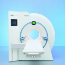
Device Information and Specification
CLINICAL APPLICATION
Whole body
GRE, IR, FIR, STIR, TrueIR/FISP, FSE, FLAIR, MT, SS-FSE, MT-SE, MTC, MSE, EPI, GMR, fat/water sat./exc.
IMAGING MODES
Single, multislice, volume study, multi angle, multi oblique
TR
2.4 msec std.; 2.0 opt.; 1.8 w/ 30mT/m at 256matrix
TE
1.1 msec std.; 0.9 opt.; 0.78 w/30 mT/m at 256matrix
178 images/sec at 256 x 256 at 100% FOV
1024 x 1024 full screen display
21 micrometer in plane, 11 micrometer optional
MAGNET WEIGHT (gantry included)
3500kg, 5000kg in operation
DIMENSION H*W*D (gantry included)
POWER REQUIREMENTS
380/400/420/440/480 V
STRENGTH
20/35 mT/m standard, 30/52 opt.
Passive, act.; 1st order std./2nd opt.
| | | |
• View the DATABASE results for 'MAGNETOM Harmony™' (2).
| | | | | | Further Reading: | Basics:
|
|
| |
| | | | | |
| |
|
Device Information and Specification CLINICAL APPLICATION Whole body GRE, IR, FIR, STIR, TrueIR/FISP, FSE, FLAIR, MT, SS-FSE, MT-SE, MTC, MSE, EPI, GMR, fat/water sat./exc. IMAGING MODES Single, multislice, volume study, multi angle, multi obliqueTR 2.4 msec std.; 2.0 opt.; 1.8 w/30 mT/m at 256matrix TE 1.1 msec std.; 0.9 opt.; 0.78 w/30 mT/m at 256matrix 178 images/sec at 256 x 256 at 100% FOV1024 x 1024 full screen display 21 micrometer in plane, 11 micrometer optional 4050kg, 5500kg in operation H*W*D 236 x 215 x 160 cm w/covers POWER REQUIREMENTS 380/400/420/440/480 V STRENGTH 20/35 mT/m standard, 30/52 opt. Passive, act.; 1st order std./2nd opt. | | | |
• View the DATABASE results for 'MAGNETOM Symphony™' (2).
| | | | | | Further Reading: | Basics:
|
|
| |
| | | Searchterm 'Meter' was also found in the following services: | | | | |
| | |
| |
|

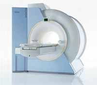
From Hitachi Medical Systems America Inc.;
the AIRIS II, an entry in the diagnostic category of open MR systems, was designed by Hitachi
Medical Systems America Inc. (Twinsburg, OH, USA) and Hitachi Medical Corp. (Tokyo) and is manufactured by the Tokyo branch. A 0.3 T field-strength magnet and phased array coils deliver high image quality without the need for a tunnel-type high-field system, thereby significantly improving patient comfort not only for claustrophobic patients.
Device Information and Specification
CLINICAL APPLICATION
Whole body
QD Head, MA Head and Neck, QD C-Spine, MA or QD Shoulder, MA CTL Spine, QD Knee, Neck, QD TMJ, QD Breast, QD Flex Body (4 sizes), Small and Large Extrem., QD Wrist, MA Foot and Ankle (WIP), PVA (WIP)
SE, GE, GR, IR, FIR, STIR, FSE, ss-FSE, FLAIR, EPI -DWI, SE-EPI, ms - EPI, SSP, MTC, SARGE, RSSG, TRSG, MRCP, Angiography: CE, 2D/3D TOF
IMAGING MODES
Single, multislice, volume study
TR
SE: 30 - 10,000msec GE: 20 - 10,000msec IR: 50 - 16,700msec FSE: 200 - 16,7000msec
TE
SE : 10 - 250msec IR: 10 -250msec GE: 5 - 50 msec FSE: 15 - 2,000
0.05 sec/image (256 x 256)
2D: 2 - 100 mm; 3D: 0.5 - 5 mm
Level Range: -2,000 to +4,000
POWER REQUIREMENTS
208/220/240 V, single phase
COOLING SYSTEM TYPE
Air-cooled
2.0 m lateral, 2.5 m vert./long
| | | |
• View the DATABASE results for 'AIRIS II™' (2).
| | | | |
| | | Searchterm 'Meter' was also found in the following services: | | | | |
| | |
| |
|

From Hitachi Medical Systems America, Inc.;
the AIRIS made its debut in 1995. Hitachi followed up with the AIRIS II system, which has proven equally successfully. 'All told, Hitachi has installed more than 1,000 MRI systems in the U.S., holding more than 17 percent of the total U.S. MRI installed base, and more than half of the installed base of open MR systems,' says Antonio Garcia, Frost and Sullivan industry research analyst.
Now Altaire employs a blend of innovative Hitachi features called VOSI™ technology, optimizing each sub-system's performance in concert with the
other sub-systems, to give the seamless mix of high-field performance
and the patient comfort, especially for claustrophobic patients, of open MR systems.
Device Information and Specification
CLINICAL APPLICATION
Whole body
DualQuad T/R Body Coil, MA Head, MA C-Spine, MA Shoulder, MA Wrist, MA CTL Spine, MA Knee, MA TMJ, MA Flex Body (3 sizes), Neck, small and large Extremity, PVA (WIP), Breast (WIP), Neurovascular (WIP), Cardiac (WIP) and MA Foot//Ankle (WIP)
SE, GE, GR, IR, FIR, STIR, ss-FSE, FSE, DE-FSE/FIR, FLAIR, ss/ms-EPI, ss/ms EPI- DWI, SSP, MTC, SE/GE-EPI, MRCP, SARGE, RSSG, TRSG, BASG, Angiography: CE, PC, 2D/3D TOF
IMAGING MODES
Single, multislice, volume study
TR
SE: 30 - 10,000msec GE: 3.6 - 10,000msec IR: 50 - 16,700msec FSE: 200 - 16,7000msec
TE
SE : 8 - 250msec IR: 5.2 -7,680msec GE: 1.8 - 2,000 msec FSE: 5.2 - 7,680
0.05 sec/image (256 x 256)
2D: 2 - 100 mm; 3D: 0.5 - 5 mm
Level Range: -2,000 to +4,000
COOLING SYSTEM TYPE
Water-cooled
3.1 m lateral, 3.6 m vertical
| | | |
• View the DATABASE results for 'Altaire™' (2).
| | | | | | Further Reading: | News & More:
|
|
| |
| | | | |
| | | |
|
| |
 | Look
Ups |
| |