 | Info
Sheets |
| | | | | | | | | | | | | | | | | | | | | | | | |
 | Out-
side |
| | | | |
|
| | | | |
Result : Searchterm 'Volume Shim' found in 1 term [ ] and 3 definitions [ ] and 3 definitions [ ], (+ 18 Boolean[ ], (+ 18 Boolean[ ] results ] results
| 1 - 5 (of 22) nextResult Pages :  [1] [1]  [2 3 4 5] [2 3 4 5] |  | |  | Searchterm 'Volume Shim' was also found in the following service: | | | | |
|  |  |
| Volume Shim |  |
| |
|
Shimming corrects local magnetic field inhomogeneities by adjusting the static gradients. A volume shim enables the shim volume to be limited. This local 3D shim provides a more precise result and therefore a better fat saturation. For spectroscopy, it provides a better starting value for an interactive shim. | |  | | | | • Share the entry 'Volume Shim':    | | | | |  Further Reading: Further Reading: | | Basics:
|
|
News & More:
| |
| |
|  | |  |  |  |
| |
|

From Hitachi Medical Systems America Inc.;
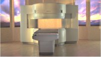
the AIRIS II, an entry in the diagnostic category of open MR systems, was designed by Hitachi
Medical Systems America Inc. (Twinsburg, OH, USA) and Hitachi Medical Corp. (Tokyo) and is manufactured by the Tokyo branch. A 0.3 T field-strength magnet and phased array coils deliver high image quality without the need for a tunnel-type high-field system, thereby significantly improving patient comfort not only for claustrophobic patients.
Device Information and Specification
CLINICAL APPLICATION
Whole body
QD Head, MA Head and Neck, QD C-Spine, MA or QD Shoulder, MA CTL Spine, QD Knee, Neck, QD TMJ, QD Breast, QD Flex Body (4 sizes), Small and Large Extrem., QD Wrist, MA Foot and Ankle (WIP), PVA (WIP)
SE, GE, GR, IR, FIR, STIR, FSE, ss-FSE, FLAIR, EPI -DWI, SE-EPI, ms - EPI, SSP, MTC, SARGE, RSSG, TRSG, MRCP, Angiography: CE, 2D/3D TOF
IMAGING MODES
Single, multislice, volume study
TR
SE: 30 - 10,000msec GE: 20 - 10,000msec IR: 50 - 16,700msec FSE: 200 - 16,7000msec
TE
SE : 10 - 250msec IR: 10 -250msec GE: 5 - 50 msec FSE: 15 - 2,000
0.05 sec/image (256 x 256)
2D: 2 - 100 mm; 3D: 0.5 - 5 mm
Level Range: -2,000 to +4,000
POWER REQUIREMENTS
208/220/240 V, single phase
COOLING SYSTEM TYPE
Air-cooled
2.0 m lateral, 2.5 m vert./long
| |  | |
• View the DATABASE results for 'AIRIS II™' (2).
| | | | |
|  | |  |  |  |
| |
|
Fat suppression is the process of utilizing specific MRI parameters to remove the deleterious effects of fat from the resulting images , e.g. with STIR, FAT SAT sequences, water selective (PROSET WATS - water only selection, also FATS - fat only selection possible) excitation techniques, or pulse sequences based on the Dixon method.
Spin magnetization can be modulated by using special RF pulses. CHESS or its variations like SPIR, SPAIR ( Spectral Selection Attenuated Inversion Recovery) and FAT SAT use frequency selective excitation pulses, which produce fat saturation.
Fat suppression techniques are nearly used in all body parts and belong to every standard MRI protocol of joints like knee, shoulder, hips, etc.

Image Guidance
Imaging of, e.g. the foot can induce bad fat suppression with SPIR/FAT SAT due to the asymmetric volume of this body part. The volume of the foot alters the magnetic field to a different degree than the smaller volume of the lower leg affecting the protons there. There is only a small band of tissue where the fat protons are precessing at the frequency expected, resulting in frequency selective fat saturation working only in that area. This can be corrected by volume shimming or creating a more symmetrical volume being imaged with water bags.
Even with their longer scan time and motion sensitivity, STIR (short T1/tau inversion recovery) sequences are often the better choice to suppress fat. STIR images are also preferred because of the decreased sensitivity to field inhomogeneities, permitting larger fields of views when compared to fat suppressed images and the ability to image away from the isocenter. See also Knee MRI.
Sequences based on Dixon turbo spin echo ( fast spin echo) can deliver a significant better fat suppression than conventional TSE/FSE imaging.
| | | |  | |
• View the DATABASE results for 'Fat Suppression' (28).
| | | | |  Further Reading: Further Reading: | Basics:
|
|
News & More:
| |
| |
|  |  | Searchterm 'Volume Shim' was also found in the following service: | | | | |
|  |  |
| |
|
Quick Overview
A disturbance of the field homogeneity, because of magnetic material (inside or outside the patient), technical problems or scanning at the edge of the field.
When images were obtained in a progression from the center to the edge of the coil, the homogeneity of the field observed by the imaged volume, changes when the distance from the center of the volume increase.
The same problem appears by scanning at a distance from the isocenter in left-right direction or too large field of view.
There are different types of bad image quality, the images are noisy, distorted or the fat suppression doesn't work because of badly set shim currents.
E.g. by using an IR sequence, changes in the T1 recovery rates of the tissues are involved. The inversion time at the center of the imaged volume is appropriate to suppress fat, but at the edge of the coil the same inversion time is sufficient to suppress water. Since the inversion time is not changed, the T1 recovery rates will increase.

Image Guidance
Take a smaller imaging volume (and for fat suppression a volume shimming), take care that the imaged region is at the center of the coil and that no magnetic material is inside the imaging volume. | |  | |
• View the DATABASE results for 'Field Inhomogeneity Artifact' (3).
| | | | |  Further Reading: Further Reading: | Basics:
|
|
| |
|  | |  |  |  |
| |
|

From GE Healthcare;
The Signa SP 0.5T™ is an open MRI magnet that is designed for use in interventional radiology and intra-operative imaging. The vertical gap configuration increases patient positioning options, improves patient observation, and allows continuous access to the patient during imaging.
The magnet enclosure also incorporates an intercom, patient observation video camera, laser patient alignment lights, and task lighting in the imaging volume.
Device Information and Specification CLINICAL APPLICATION Whole body Integrated transmit and receive body coil; optional rotational body coil, head; other coils optional; open architecture makes system compatible with a wide selection of coilsarray Standard: SE, IR, 2D/3D GRE and SPGR, 2D/3D TOF, 2D/3D FSE, 2D/3D FGRE and FSPGR, SSFP, FLAIR, EPI, optional: 2D/3D Fiesta, true chem sat, fat/water separation, single shot diffusion EPI IMAGING MODES Localizer, single slice, multislice, volume, fast, POMP, multi slab, cine, slice and frequency zip, extended dynamic range, tailored RF TR 1.3 to 12000 msec in increments of 1 msec TE 0.4 to 2000 msec in increments of 1 msec 2D: 1.4mm - 20mm 3D: 0.2mm - 20mm POWER REQUIREMENTS 200 - 480, 3-phase | |  | |
• View the DATABASE results for 'Signa SP 0.5T™ Open Configuration' (2).
| | | | |  Further Reading: Further Reading: | News & More:
|
|
| |
|  | |  |  |
|  | 1 - 5 (of 22) nextResult Pages :  [1] [1]  [2 3 4 5] [2 3 4 5] |
| |
|
| |
 | Look
Ups |
| |