| Info
Sheets |
| | | | | | | | | | | | | | | | | | | | | | | | |
 | Out-
side |
| | | | |
|
| | | 'MR Guided Interventions' | |
Result : Searchterm 'MR Guided Interventions' found in 1 term [ ] and 7 definitions [ ] and 7 definitions [ ], (+ 1 Boolean[ ], (+ 1 Boolean[ ] results ] results
| | previous 6 - 9 (of 9) Result Pages : [1] [2] |  | |  | Searchterm 'MR Guided Interventions' was also found in the following services: | | | | |
| |  |
| |
|
| | | | | | | | | | |  Further Reading: Further Reading: | | Basics:
|
|
News & More:
|  |
Safety of Bedside Portable Low-Field Brain MRI in ECMO Patients Supported on Intra-Aortic Balloon Pump
Friday, 18 November 2022 by www.mdpi.com | | |
Researchers at the University of Tsukuba develop a portable MRI system specifically for identifying wrist cartilage damage among athletes, providing a convenient means of early detection and treatment of injuries
Tuesday, 26 April 2022 by www.tsukuba.ac.jp | | |
This bizarre looking helmet can create better brain scans
Friday, 11 February 2022 by www.sciencedaily.com | | |
A low-cost and shielding-free ultra-low-field brain MRI scanner
Tuesday, 14 December 2021 by www.nature.com | | |
Portable MRI provides life-saving information to doctors treating strokes
Thursday, 5 August 2021 by news.yale.edu | | |
Synaptive Evry, an MRI for Any Space, Cleared by FDA
Thursday, 30 April 2020 by www.medgadget.com | | |
World's First Portable MRI Cleared by FDA
Monday, 17 February 2020 by www.medgadget.com | | |
Introducing a point-of-care MRI system
Tuesday, 29 October 2019 by healthcare-in-europe.com | | |
Opportunities in Interventional and Diagnostic Imaging by Using High-performance Low-Field-Strength MRI
Tuesday, 1 October 2019 by pubs.rsna.org | | |
Portable 'battlefield MRI' comes out of the lab
Thursday, 30 April 2015 by physicsworld.com | | |
Portable MRI could aid wounded soldiers and children in the third world
Thursday, 23 April 2015 by phys.org |
|
| |
| | | | | |
| |
|

It is important to remember when working around a superconducting magnet that the magnetic field is always on. Under usual working conditions the field is never turned off. Attention must be paid to keep all ferromagnetic items at an adequate distance from the magnet. Ferromagnetic objects which came accidentally under the influence of these strong magnets can injure or kill individuals in or nearby the magnet, or can seriously damage every hardware, the magnet itself, the cooling system, etc..
See MRI resources Accidents.
The doors leading to a magnet room should be closed at all times except when entering or exiting the room. Every person working in or entering the magnet room or adjacent rooms with a magnetic field has to be instructed about the dangers. This should include the patient, intensive-care staff, and maintenance-, service- and cleaning personnel, etc..
The 5 Gauss limit defines the 'safe' level of static magnetic field exposure. The value of the absorbed dose is fixed by the authorities to avoid heating of the patient's tissue and is defined by the specific absorption rate.
Leads or wires that are used in the magnet bore during imaging procedures, should not form large-radius wire loops. Leg-to-leg and leg-to-arm skin contact should be prevented in order to avoid the risk of burning due to the generation of high current loops if the legs or arms are allowed to touch. The patient's skin should not be in contact with the inner bore of the magnet.
The outflow from cryogens like liquid helium is improbable during normal operation and not a real danger for patients.
The safety of MRI contrast agents is tested in drug trials and they have a high compatibility with very few side effects. The variations of the side effects and possible contraindications are similar to X-ray contrast medium, but very rare. In general, an adverse reaction increases with the quantity of the MRI contrast medium and also with the osmolarity of the compound.
See also 5 Gauss Fringe Field, 5 Gauss Line, Cardiac Risks, Cardiac Stent, dB/dt, Legal Requirements, Low Field MRI, Magnetohydrodynamic Effect, MR Compatibility, MR Guided Interventions, Claustrophobia, MRI Risks and Shielding. | | | | | | | | |
• View the DATABASE results for 'MRI Safety' (42).
| | |
• View the NEWS results for 'MRI Safety' (13).
| | | | | | Further Reading: | Basics:
|
|
News & More:
| |
| |
| | | | | |
| |
|
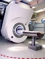
Device Information and Specification
CLINICAL APPLICATION
Whole body
CONFIGURATION
Mobile compact
Whole body, intra-operative head, neck volume, atlas head//neck vascular quadrature phased array, spine quadrature, C/T/L spine phased array, small joint, large joint, TMJ bilateral, shoulder phased array, extremity quadrature volume, wrist, hand quadrature, general purpose flexible, pelvis/abdomen phased array, body quadrature, phased array flexible, breast bilateral
IMAGING MODES
Localizer, single slice, multislice, volume
| | | |
• View the DATABASE results for 'iMotion™ 1.5 Tesla Magnet' (2).
| | | | |
| | | Searchterm 'MR Guided Interventions' was also found in the following services: | | | | |
| |  |
| |
|

From Philips Medical Systems;
the Panorama 0.23 T, providing a new design optimized for patient comfort, faster reconstruction time than before (300 images/second) and new gradient
specifications. Philips' Panorama 0.23 T I/T supports MR- guided interventions, resulting in minimally invasive procedures, more targeted surgery, reduced recovery time and shorter hospital stays. Optional OptoGuide functionality enables real-time needle tracking. Philips' Panorama 0.23 TPanorama 0.2 R/T is the first and only open MRI system to enable radiation therapy planning using MR data sets. The Panorama also features the new and consistent Philips User Interface, an essential element of the Vequion clinical IT family of products and services.
Device Information and Specification CLINICAL APPLICATION Whole body SE, FE, IR, FFE, DEFFE, DESE, TSE, DETSE, Single shot SE, DRIVE, Balanced FFE, MRCP, Fluid Attenuated Inversion Recovery, Turbo FLAIR, IR-TSE, T1-STIR TSE, T2-STIR TSE, Diffusion Imaging, 3D SE, 3D FFE, MTC;; Angiography: CE-ANGIO, MRA 2D, 3D TOFOpen x 46 cm x infinite (side-first patient entry) POWER REQUIREMENTS 400/480 V COOLING SYSTEM TYPE Closed loop chilled water ( chiller included) | | | |
• View the DATABASE results for 'Panorama 0.23T™' (2).
| | | | | | Further Reading: | News & More:
|
|
| |
| | | | |
| | | |
|
| |
 | Look
Ups |
| |