| Info
Sheets |
| | | | | | | | | | | | | | | | | | | | | | | | |
 | Out-
side |
| | | | |
|
| | | | |
Result : Searchterm 'Oblique Imaging' found in 1 term [ ] and 0 definition [ ] and 0 definition [ ], (+ 16 Boolean[ ], (+ 16 Boolean[ ] results ] results
| | previous 11 - 15 (of 17) nextResult Pages : [1] [2 3 4] |  | | | | |  |
| |
|
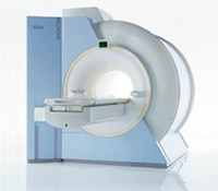
Device Information and Specification CLINICAL APPLICATION Whole body GRE, IR, FIR, STIR, TrueIR/FISP, FSE, FLAIR, MT, SS-FSE, MT-SE, MTC, MSE, EPI, GMR, fat/water sat./exc. IMAGING MODES Single, multislice, volume study, multi angle, multi obliqueTR 2.4 msec std.; 2.0 opt.; 1.8 w/30 mT/m at 256matrix TE 1.1 msec std.; 0.9 opt.; 0.78 w/30 mT/m at 256matrix 178 images/sec at 256 x 256 at 100% FOV1024 x 1024 full screen display 21 micrometer in plane, 11 micrometer optional 4050kg, 5500kg in operation H*W*D 236 x 215 x 160 cm w/covers POWER REQUIREMENTS 380/400/420/440/480 V STRENGTH 20/35 mT/m standard, 30/52 opt. Passive, act.; 1st order std./2nd opt. | | | | | | | | | | |  Further Reading: Further Reading: | Basics:
|
|
| |
| | | | | |
| |
|
In the last years, cardiac MRI techniques have progressively improved. No other noninvasive imaging modality provides the same degree of contrast and temporal resolution for the assessment of cardiovascular anatomy and pathology. Contraindications MRI are the same as for other magnetic resonance techniques.
The primary advantage of MRI is extremely high contrast resolution between different tissue types, including blood. Moreover, MRI is a true 3 dimensional imaging modality and images can be obtained in any oblique plane along the true cardiac axes while preserving high temporal and spatial resolution with precise demonstration of cardiac anatomy without the administration of contrast media.
Due to these properties, MRI can precisely characterize cardiac function and quantify cavity volumes, ejection fraction, and left ventricular mass. In addition, cardiac MRI has the ability to quantify flow (see flow quantification), including bulk flow in vessels, pressure gradients across stenosis, regurgitant fractions and shunt fractions. Valve morphology and area can be determined and the severity of stenosis quantified. In certain disease states, such as myocardial infarction, the contrast resolution of MRI is further improved by the addition of extrinsic contrast agents (see myocardial late enhancement).
A dedicated cardiac coil, and a field strength higher than 1 Tesla is recommended to have sufficient signal. Cardiac MRI acquires ECG gating. Cardiac gating (ECGs) obtained within the MRI scanner, can be degraded by the superimposed electrical potential of flowing blood in the magnetic field. Therefore, excellent contact between the skin and ECG leads is necessary. For male patients, the skin at the lead sites can be shaved. A good cooperation of the patient is necessary because breath holding at the end of expiration is practiced during the most sequences.
See also Displacement Encoding with Stimulated Echoes.
For Ultrasound Imaging (USI) see Cardiac Ultrasound at Medical-Ultrasound-Imaging.com.
See also the related poll results: ' In 2010 your scanner will probably work with a field strength of' and ' MRI will have replaced 50% of x-ray exams by' | | | | | |
• View the DATABASE results for 'Cardiac MRI' (15).
| | |
• View the NEWS results for 'Cardiac MRI' (15).
| | | | | | Further Reading: | | Basics:
|
|
News & More:
|  |
MRI technology visualizes heart metabolism in real time
Friday, 18 November 2022 by medicalxpress.com | | |
Even early forms of liver disease affect heart health, Cedars-Sinai study finds
Thursday, 8 December 2022 by www.eurekalert.org | | |
MRI sheds light on COVID vaccine-associated heart muscle injury
Tuesday, 15 February 2022 by www.sciencedaily.com | | |
Radiologists must master cardiac CT, MRI to keep pace with demand: The heart is not a magical organ
Monday, 1 March 2021 by www.radiologybusiness.com | | |
Diffusion weighted imaging (DWI) and diffusion tensor imaging (DTI) in the heart (myocardium)
Sunday, 30 August 2020 by github.com | | |
Non-invasive diagnostic procedures for suspected CHD: Search reveals informative evidence
Wednesday, 8 July 2020 by medicalxpress.co | | |
Cardiac MRI Becoming More Widely Available Thanks to AI and Reduced Exam Times
Wednesday, 19 February 2020 by www.dicardiology.com | | |
Controlling patient's breathing makes cardiac MRI more accurate
Friday, 13 May 2016 by www.upi.com | | |
Precise visualization of myocardial injury: World's first patient-based cardiac MRI study using 7T MRI
Wednesday, 10 February 2016 by medicalxpress.com | | |
New technique could allow for safer, more accurate heart scans
Thursday, 10 December 2015 by www.gizmag.com |
|
| |
| | | | | |
| |
|
Magnetic resonance imaging ( MRI) of the spine is a noninvasive procedure to evaluate different types of tissue, including the spinal cord, vertebral disks and spaces between the vertebrae through which the nerves travel, as well as distinguish healthy tissue from diseased tissue.
The cervical, thoracic and lumbar spine MRI should be scanned in individual sections.
The scan protocol parameter like e.g. the field of view ( FOV), slice thickness and matrix are usually different for cervical, thoracic and lumbar spine MRI, but the method
is similar. The standard views in the basic spinal MRI scan to create detailed slices (cross sections) are sagittal T1 weighted and T2 weighted images over the whole body part, and transverse (e.g. multi angle oblique) over the region of interest with different pulse sequences according to the result of the sagittal slices. Additional views or different types of pulse sequences like fat suppression, fluid attenuation inversion recovery ( FLAIR) or
diffusion weighted imaging are created dependent on the indication.
Indications:
•
Neurological deficit, evidence of radiculopathy, cauda equina compression
•
Primary tumors or drop metastases
•
Infection/inflammatory disease, multiple sclerosis
•
Postoperative evaluation of lumbar spine: disk vs. scar
•
Localized back pain with no radiculopathy (leg pain)
Contrast enhanced MRI techniques delineate infections vs. malignancies, show a syrinx cavity and support to differentiate the postoperative conditions. After surgery for disk disease, significant fibrosis can occur in the spine. This scarring can mimic residual disk herniation. Magnetic resonance myelography evaluates spinal stenosis and various intervertebral discs can be imaged with multi angle oblique techniques. Cine series can be used to show true range of motion studies of parts of the spine.
Advanced open MRI devices are developed to perform positional scans in the position of pain or symptom (e.g. Upright™ MRI formerly Stand-Up MRI). | | | | | |
• View the DATABASE results for 'Spine MRI' (11).
| | |
• View the NEWS results for 'Spine MRI' (4).
| | | | | | Further Reading: | | Basics:
|
|
News & More:
| |
| |
| | | | | |
| |
|
MRI of the lumbar spine, with its multiplanar 3 dimensional imaging capability, is currently the preferred modality for establishing a diagnosis. MRI scans and magnetic resonance myelography have many advantages compared with computed tomography and/or X-ray myelography in evaluating the lumbar spine. MR imaging scans large areas of the spine without ionizing radiation, is noninvasive, not affected by bone artifacts, provides vascular imaging capability, and makes use of safer contrast agents ( gadolinium chelate).
Due to the high level of tissue contrast resolution, nerves and discs are clearly visible. MRI is excellent for detecting degenerative disease in the spine. Lumbar spine MRI accurately shows disc disease (prolapsed disc or slipped disc), the level at which disc disease occurs, and if a disc is compressing spinal nerves. Lumbar spine MRI depicts soft tissues, including the cauda equina, spinal cord, ligaments, epidural fat, subarachnoid space, and intervertebral discs. Loss of epidural fat on T1 weighted images, loss of cerebrospinal fluid signal around the dural sac on T2 weighted images and degenerative disc disease are common features of lumbar stenosis.
Common indications for MRI of the lumbar spine:
•
Neurologic deficits, evidence of radiculopathy, acute spinal cord compression (e.g., sudden bowel/bladder disturbance)
•
Suspected systemic disorders (primary tumors, drop metastases, osteomyelitis)
•
Postoperative evaluation of lumbar spine: disk vs. scar
•
Localized back pain with no radiculopathy (leg pain)
Lumbar spine imaging requires a special spine coil. often used whole spine array coils have the advantage that patients do not need other positioning if also upper parts of the spine should be scanned. Sagittal T1 and T2 weighted FSE sequences are the standard views. With multi angle oblique techniques individually oriented transverse images of each intervertebral disc at different angles can be obtained.
See also the related poll result: ' MRI will have replaced 50% of x-ray exams by' | | | | | |
• View the DATABASE results for 'Lumbar Spine MRI' (6).
| | | | | | Further Reading: | Basics:
|
|
News & More:
| |
| |
| | | | | |
| |
|
MRI of the shoulder with its excellent soft tissue discrimination, and high spatial resolution offers the best noninvasive way to study the shoulder. MRI images of the bone, muscles and tendons of the glenohumeral joint can be obtained in any oblique planes and projections. MRI gives excellent depiction of rotator cuff tears, injuries to the biceps tendon and damage to the glenoid labrum. Shoulder MRI is better than ultrasound imaging at depicting structural changes such as osteophytic spurs, ligament thickening, and acromial shape that may have predisposed to tendon degeneration.
A dedicated shoulder coil and careful patient positioning in external rotation with the shoulder as close as reasonably possible to the center of the magnet is necessary for a good image quality. If possible, the opposite shoulder should be lifted up, so that the patient lies on the imaged shoulder in order to rotate and fix this shoulder to reduce motion during breathing.
Axial, coronal oblique, and sagittal oblique proton density with fat suppression, T2 and T1 provide an assessment of the rotator cuff, biceps, deltoid, acromio-clavicular joint, the glenohumeral joint and surrounding large structures. If a labral injury is suspected, a Fat Sat gradient echo sequence is recommended. In some cases, a direct MR shoulder arthrogram with intra-articular injection of dilute gadolinium or an indirect arthrogram with imaging 20 min. after intravenous injection may be helpful. See also Imaging of the Extremities. | | | | | | | | | | |
• View the DATABASE results for 'Shoulder MRI' (3).
| | |
• View the NEWS results for 'Shoulder MRI' (1).
| | | | | | Further Reading: | News & More:
|
|
| |
| | | | |
| | | |
|
| |
 | Look
Ups |
| |