 | Info
Sheets |
| | | | | | | | | | | | | | | | | | | | | | | | |
 | Out-
side |
| | | | |
|
| | | | | |  | Searchterm 'cardiac' was also found in the following services: | | | | |
|  |  |
| |
|
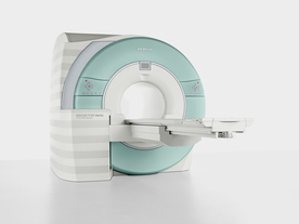
From Siemens Medical Systems;
Received FDA clearance in 2007.
The MAGNETOM Verio provides up to 102 integrated matrix coil elements and up to 32 independent radiofrequency channels that allow flexible coil combinations to make patient and coil repositioning virtually unnecessary. The Tim (total imaging matrix) technology also increases patient throughput due to a shorter scan time.
The open bore design offers great comfort for patients of all shapes and sizes.
Device Information and Specification
CLINICAL APPLICATION
Whole Body
CONFIGURATION
Ultra-short open bore
Head, spine, torso/ body coil, neurovascular, cardiac, neck and multi-purpose flex coils. Peripheral vascular, breast, shoulder, knee, wrist, foot//ankle, TMJ optional.
CHANNELS (min. / max. configuration)
8, 18, 32
Chemical shift imaging, single voxel spectroscopy
MAGNET WEIGHT (gantry included)
8200 kg
DIMENSION H*W*D (gantry included)
173 x 230 x 222 cm
Passive, active; first order,
second order standard
POWER REQUIREMENTS
380 / 400 / 420 / 440 / 460 / 480 V, 3-phase + ground; 110 kVA
| | | | | |
| | | | | |
| |
|
The MRI device is located within a specially shielded room ( Faraday cage) to avoid outside interference, caused by the use of radio waves very close in frequency to those of ordinary FM radio stations.
The MRI procedure can easily be performed through clothing and bones, but attention must be paid to ferromagnetic items, because they will be attracted from the magnetic field. A hospital gown is appropriate, or the patient should wear clothing without metal fasteners and remove any metallic objects like hairpins, jewelry, eyeglasses, clocks, hearing aids, any removable dental work, lighters, coins etc., not only for MRI safety reasons.
Metal in or around the scanned area can also cause errors in the reconstructed images ( artifacts). Because the strong magnetic field can displace, or disrupt metallic objects, people with an implanted active device like a cardiac pacemaker cannot be scanned under normal circumstances and should not enter the MRI area.
The MRI machine can look like a short tunnel or has an open MRI design and the magnet does not completely surround the patient. Usually the patient lies on a comfortable motorized table, which slides into the scanner, depending on the MRI device, patients may be also able to sit up. If a contrast agent is to be administered, intravenous access will be placed. A technologist will operate the MRI machine and observe the patient during the examination from an adjacent room. Several sets of images are usually required, each taking some minutes. A typical MRI scan includes three to nine imaging sequences and may take up to one hour. Improved MRI devices with powerful magnets, newer software, and advanced sequences may complete the process in less time and better image quality.
Before and after the most MRI procedures no special preparation, diet, reduced activity, and extra medication is necessary. The magnetic field and radio waves are not felt and no pain is to expect.
Movement can blur MRI images and cause certain artifacts. A possible problem is the claustrophobia that some patients experience from being inside a tunnel-like scanner. If someone is very anxious or has difficulty to lie still, a sedative agent may be given. Earplugs and/or headphones are usually given to the patient to reduce the loud acoustic noise, which the machine produces during normal operation. A technologist observes the patient during the test. Some MRI scanners are equipped with televisions and music to help the examination time pass.
MRI is not a cheap examination, however cost effective by eliminating the need for invasive radiographic procedures, biopsies, and exploratory surgery. MRI scans can also save money while minimizing patient risk and discomfort. For example, MRI can reduce the need for X-ray angiography and myelography, and can eliminate unnecessary diagnostic procedures that miss occult disease. See also Magnetic Resonance Imaging MRI, Medical Imaging, Cervical Spine MRI, Claustrophobia, MRI Risks and Pregnancy.
For Ultrasound Imaging (USI) see Ultrasound Imaging Procedures at Medical-Ultrasound-Imaging.com.
See also the related poll result: ' MRI will have replaced 50% of x-ray exams by' | | | | | |
• View the DATABASE results for 'MRI Procedure' (11).
| | |
• View the NEWS results for 'MRI Procedure' (6).
| | | | |  Further Reading: Further Reading: | News & More:
|
|
| |
| | | | | |
| |
|
The subacute risks and side effects of magnetic and RF fields (for patients and staff) have been intensively examined for a long time, but there have been no long-term studies following persons who have been exposed to the static magnetic fields used in MRI. However, no permanent hazardous effects of a static magnetic field exposure upon human beings have yet been demonstrated.
Temporary possible side effects of high magnetic and RF fields:
•
Varying magnetic fields can induce so-called magnetic phosphenes that occur when an individual is subject to rapid changes of 2-5 T/s, which can produce a flashing sensation in the eyes. This temporary side effect does not seem to damage the eyes. Static field strengths used for clinical MRI examinations vary between 0.2 and 3.0 tesla;; field changes during the MRI scan vary in the dimension of mT/s. Experimental imaging units can use higher field strengths of up to 14.0 T, which are not approved for human use.
•
The Radio frequency pulses mainly produce heat, which is absorbed by the body tissue. If the power of the RF radiation is very high, the patient may be heated too much. To avoid this heating, the limit of RF exposure in MRI is up to the maximum specific absorption rate (SAR) of 4 W/kg whole body weight (can be different from country to country). For MRI safety reasons, the MRI machine starts no sequence, if the SAR limit is exceeded.
•
Very high static magnetic fields are needed to reduce the conductivity of nerves perceptibly. Augmentation of T waves is observed at fields used in standard imaging but this side effect in MRI is completely reversible upon removal from the magnet. Cardiac arrhythmia threshold is typically set to 7-10 tesla. The magnetohydrodynamic effect, which results from a voltage occurring across a vessel in a magnetic field and percolated by a saline solution such as blood, is irrelevant at the field strengths used.
The results of some animal and cellular studies suggest the possibility that electromagnetic fields may act as co-carcinogens or tumor promoters, but the data are inconclusive.
Up to 45 tesla, no important effects on enzyme systems have been observed. Neither changes in enzyme kinetics, nor orientation changes in macromolecules have been conclusively demonstrated.
There are some publications associating an increase in the incidence of leukemia with the location of buildings close to high-current power lines with extremely low-frequency (ELF) electromagnetic radiation of 50-60 Hz, and industrial exposure to electric and magnetic fields but a transposition of such effects to MRI or MRS seems unlikely.
Under consideration of the MRI safety guidelines, real dangers or risks of an exposure with common MRI field strengths up to 3 tesla as well as the RF exposure during the MRI scan, are not to be expected.
For more MRI safety information see also Nerve Conductivity,
Contraindications, Pregnancy
and Specific Absorption Rate.
See also the related poll result: ' In 2010 your scanner will probably work with a field strength of' | | | |
• View the DATABASE results for 'MRI Risks' (9).
| | |
• View the NEWS results for 'MRI Risks' (3).
| | | | | | Further Reading: | | Basics:
|
|
News & More:
| |
| |
| | | Searchterm 'cardiac' was also found in the following services: | | | | |
| | |
| |
|
The definition of a scan is to form an image or an electronic representation. The MRI scan uses magnetic resonance principles to produce extremely detailed pictures of the body tissue without the need for X-ray exposure or other damaging forms of radiation.
MRI scans show structures of the different tissues in the body. The tissue that has the least hydrogen atoms (e.g., bones) appears dark, while the tissue with many hydrogen atoms (e.g., fat) looks bright. The MRI pictures of the brain show details and abnormal structures ( brain MRI), for example, tumors, multiple sclerosis lesions, bleedings, or brain tissue that has suffered lack of oxygen after a stroke.
A cardiac MRI scan demonstrates the heart as well as blood vessels ( cardiovascular imaging) and is used to detect heart defects with e.g., changes in the thickness and infarctions of the muscles around the heart. With MRI scans, nearly all kind of body parts can be tested, for example the joints like knee and shoulder, lumbar, thoracic and cervical spine, the pelvis including fetal MRI, and the soft parts of the body such as the liver, kidneys, and spleen.
The MRI procedure includes three to nine imaging sequences and may take up to one hour. See also Lumbar Spine MRI, MRI Safety and Open MRI. | | | | | | | | | | |
• View the DATABASE results for 'MRI Scan' (31).
| | |
• View the NEWS results for 'MRI Scan' (95).
| | | | | | Further Reading: | | Basics:
|
|
News & More:
|  |
A Knee MRI in Half the Time? It's Possible
Thursday, 8 April 2021 by www.diagnosticimaging.com | | |
Michigan radiologist warns about 'incidental findings' in full body MRI scans
Wednesday, 4 October 2023 by www.wilx.com | | |
ACCELERATING MRI SCANS WITH ARTIFICIAL INTELLIGENCE
Friday, 28 August 2020 by www.analyticsinsight.net | | |
Radiographer's Lego Open MRI Product Idea Reaches New Milestone
Monday, 11 November 2019 by www.itnonline.com | | |
Why we need erasable MRI scans
Wednesday, 25 April 2018 by phys.org | | |
MRI as accurate as CT for Crohn's disease detection, management
Tuesday, 6 June 2017 by www.healthimaging.com | | |
MRI scans predict patients' ability to fight the spread of cancer
Tuesday, 12 December 2017 by eurekalert.org | | |
Audio/Video System helps patients relax during MRI scans
Monday, 8 December 2014 by news.thomasnet.com | | |
MRI scans could be a 'game-changer' in prostate cancer testing
Tuesday, 5 August 2014 by www.abc.net.au | | |
7-Tesla MRI scanner allows even more accurate diagnosis of breast cancer
Thursday, 6 March 2014 by www.healthcanal.com |
|
| |
| | | | | | | | | |
| | | |
|
| |
 | Look
Ups |
| |


