Magnetic Resonance - Technology Information Portal Welcome to MRI Technology
Info
  Sheets

Out-
      side


 'Voxel' 
SEARCH FOR    
 
  2 3 5 A B C D E F G H I J K L M N O P Q R S T U V W X Y Z
Result : Searchterm 'Voxel' found in 4 terms [] and 35 definitions []
previous     36 - 39 (of 39)     
Result Pages : [1]  [2 3 4 5 6 7 8]
Searchterm 'Voxel' was also found in the following services: 
spacer
News  (1)  Resources  (1)  
 
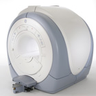
Signa HDx 3.0T™InfoSheet: - Devices -
Intro, 
Types of Magnets, 
Overview, 
etc.
 
gehealthcare.com/euen/mri/products/signa-hdx-3t/index.html From GE Healthcare;
The Signa HDx MRI system is GE's leading edge whole body magnetic resonance scanner designed to support high resolution, high signal to noise ratio, and short scan times.
Signa HDx 3.0T offers new technologies like ultra-fast image reconstruction through the new XVRE recon engine, advancements in parallel imaging algorithms and the broadest range of premium applications. The HD applications, PROPELLER (high-quality brain imaging extremely resistant to motion artifacts), TRICKS (contrast-enhanced angiographic vascular lower leg imaging), VIBRANT (for breast MRI), LAVA (high resolution liver imaging with shorter breath holds and better organ coverage) and MR Echo (high-definition cardiac images in real time) offer unique capabilities.
Device Information and Specification
CLINICAL APPLICATION
Whole body
CONFIGURATION
Compact short bore
Head and body coil, T/R quadrature head; optional coils e.g., T/R phased array extremity abdomen, spine, breast, knee, shoulder, cardiac imaging coils
SYNCHRONIZATION
ECG/peripheral, respiratory gating
PULSE SEQUENCES
SE, IR, 2D/3D GRE, RF-spoiled GRE, 2DFGRE, 2DFSPGR, 3DFGRE, 3DFSPGR, 3DTOFGRE, 3DFSPGR, 2DFSE, 2DFSE-XL, 2DFSE-IR, T1-FLAIR, SSFSE, EPI, DW-EPI, BRAVO, Angiography: 2D/3D TOF, 2D/3D phase contrast vascular
IMAGING MODES
Single, multislice, volume study, fast scan, multi slab, cine, localizer
1 cm to 40 cm continuous
2D 0.5 mm; 3D 0.1 mm
1024 x 1024
PIXEL INTENSITY
256 gray levels
60 cm
MAGNET WEIGHT
12000 kg
H*W*D
240 x 2216,6 x 201,6 cm
POWER REQUIREMENTS
480 or 380/415, 3 phase ||
COOLING SYSTEM TYPE
Closed-loop water-cooled grad.
0.03 L/hr helium
STRENGTH
23 - 50 mT/m
80 - 150 mT/m/ms
5-GAUSS FRINGE FIELD
2.8 m / 5.0 m
second and high order
spacer
Searchterm 'Voxel' was also found in the following services: 
spacer
Radiology  (5) Open this link in a new windowUltrasound  (2) Open this link in a new window
Signal to Noise Ratio
 
(SNR or S/N) The signal to noise ratio is used in MRI to describe the relative contributions to a detected signal of the true signal and random superimposed signals ('background noise') - a criterion for image quality.
One common method to increase the SNR is to average several measurements of the signal, on the expectation that random contributions will tend to cancel out. The SNR can also be improved by sampling larger volumes (increasing the field of view and slice thickness with a corresponding loss of spatial resolution) or, within limits, by increasing the strength of the magnetic field used. Surface coils can also be used to improve local signal intensity. The SNR will depend, in part, on the electrical properties of the sample or patient being studied. The SNR increases in proportion to voxel volume (1/resolution), the square root of the number of acquisitions (NEX), and the square root of the number of scans (phase encodings). SNR decreases with the field of view squared (FOV2) and wider bandwidths. See also Signal Intensity and Spin Density.

Measuring SNR:
Record the mean value of a small ROI placed in the most homogeneous area of tissue with high signal intensity (e.g. white matter in thalamus). Calculate the standard deviation for the largest possible ROI placed outside the object in the image background (avoid ghosting/aliasing or eye movement artifact regions).
The SNR is then:
Mean Signal/Standard Deviation of Background Noise
 
Images, Movies, Sliders:
 Brain MRI Images T1  Open this link in a new window


spacer

• View the DATABASE results for 'Signal to Noise Ratio' (48).Open this link in a new window


• View the NEWS results for 'Signal to Noise Ratio' (2).Open this link in a new window.
 
Further Reading:
  Basics:
Musculoskeletal MRI at 3.0 T: Relaxation Times and Image Contrast
Sunday, 1 August 2004   by www.ajronline.org    
  News & More:
Picture-Perfect Particles Enhance MRI Signal
Friday, 13 April 2012   by cen.acs.org    
Researchers design 'intelligent' metamaterial to make MRIs affordable and accessible
Tuesday, 5 November 2019   by phys.org    
Metamaterials boost sensitivity of MRI machines
Thursday, 14 January 2016   by www.eurekalert.org    
Optimizing Musculoskeletal MR
   by rad.usuhs.mil    
MRI Resources 
Abdominal Imaging - Stimulator pool - Libraries - Nerve Stimulator - RIS - Patient Information
 
Tomographic Imaging
 
Tomography is imaging by sections or sectioning. A device used in tomography is called a tomograph, while the image produced is a tomogram. The mathematical basis for tomographic imaging was laid down by Johann Radon. It is applied in computed tomography and magnetic resonance tomography (MRT) also called magnetic resonance imaging (MRI) to obtain cross-sectional images of slices through the body of patients. Each of that slices is defined by thickness and spatial resolution (see voxel).
 
Images, Movies, Sliders:
 MRI - Anatomic Imaging of the Ankle 3  Open this link in a new window
    
SlidersSliders Overview

 Anatomic MRI of the Knee 2  Open this link in a new window
    
SlidersSliders Overview

 
spacer

• View the DATABASE results for 'Tomographic Imaging' (9).Open this link in a new window

 
Further Reading:
  Basics:
Tomographic Image Reconstruction
   by www.aapm.org    
Image Characteristics and Quality
   by www.sprawls.org    
Magnetic Resonance Imaging, History & Introduction
2000   by www.cis.rit.edu    
  News & More:
Magnetic resonance imaging
   by www.scholarpedia.org    
PET/CT Combination Can Highly Increase Detection Of Colorectal Cancer
Monday, 29 October 2007   by www.sciencedaily.com    
Searchterm 'Voxel' was also found in the following services: 
spacer
News  (1)  Resources  (1)  
 
Volumetric Imaging
 
Volumetric imaging is a 3D technique where all the MRI signals are collected from the entire tissue sample and imaged as a whole entity, therefore providing a high signal to noise ratio. The acquisition of isotropic voxels or thin slices with high spatial resolution allows to create multiplanar reconstructions in all planes; a compensation for the usually longer scan time. The acquisition time can be reduced by parallel imaging technique.
New T2 weighted variants of 3D sequences (FSE-XETA, T2-SPACE, VISTA) have been introduced that differ from conventional FSE sequences. An echo train containing up to 200 echoes obtained at a minimum echo spacing allows very fast acquisition. A flip angle modulation (flip angle sweep - FAS) during the FSE readout carries magnetization as long as possible to avoid blurring and provide optimal signal at the effective echo time. This type of imaging is well suited for brain and spine MRI procedures.
Newer T1 weighted variants include Liver Acquisition with Volume Acquisition (LAVA) and T1W High Resolution Isotropic Volume Examination (THRIVE), which have advantages for dynamic breath hold imaging in liver and abdominal examinations.

See also Volume Imaging, 3 Dimensional Imaging.
 
Images, Movies, Sliders:
 Brain MRI Sagittal T1 001  Open this link in a new window
    
 Circle of Willis, Time of Flight, MIP  Open this link in a new window
    
SlidersSliders Overview

 MRI of the Skull Base  Open this link in a new window
    
SlidersSliders Overview

 
spacer

• View the DATABASE results for 'Volumetric Imaging' (4).Open this link in a new window


• View the NEWS results for 'Volumetric Imaging' (1).Open this link in a new window.
 
Further Reading:
  Basics:
Cutting Edge Imaging of THE Spine
February 2007   by www.pubmedcentral.nih.gov    
3-D VOLUMETRIC IMAGING FOR STEREOTACTIC LESIONAL AND DEEP BRAIN STIMULATION SURGERY
Searchterm 'Voxel' was also found in the following services: 
spacer
Radiology  (5) Open this link in a new windowUltrasound  (2) Open this link in a new window
previous      36 - 39 (of 39)     
Result Pages : [1]  [2 3 4 5 6 7 8]
 Random Page
 
Share This Page
FacebookTwitterLinkedIn

MR-TIP    
Community   
User
Pass
Forgot your UserID/Password ?    


MRI is trending to low field magnets :
reduced costs will lead to this change 
AI will close the gap to high field 
only in remote areas 
is only temporary 
never 

Look
      Ups


MR-TIP.com uses cookies! By browsing MR-TIP.com, you agree to our use of cookies.

Magnetic Resonance - Technology Information Portal
Member of SoftWays' Medical Imaging Group - MR-TIP • Radiology-TIP • Medical-Ultrasound-Imaging • 
Copyright © 2003 - 2024 SoftWays. All rights reserved. [ 26 April 2024]
Terms of Use | Privacy Policy | Advertising
 [last update: 2024-02-26 03:41:00]