 | Info
Sheets |
| | | | | | | | | | | | | | | | | | | | | | | | |
 | Out-
side |
| | | | |
|
| | | | |
Result : Searchterm 'Fast Multi Planar IR' found in 0 term [ ] and 0 definition [ ] and 0 definition [ ], (+ 6 Boolean[ ], (+ 6 Boolean[ ] results ] results
| | 1 - 5 (of 6) nextResult Pages : [1 2] |  | | | | |  |
| |
|
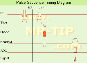
In simple ultra fast GRE imaging, TR and TE are so short, that tissues have a poor imaging signal and - more importantly - poor contrast except when contrast media enhanced ( contrast enhanced angiography). Therefore, the magnetization is 'prepared' during the preparation module, most frequently by an initial 180° inversion pulse.
In the pulse sequence timing diagram, the basic ultra fast gradient echo sequence is illustrated. The 180° inversion pulse is executed one time (to the left of the vertical line), the right side represents the data collection period and is often repeated depending on the acquisition parameters.
See also Pulse Sequence Timing Diagram, there you will find a description of the components.
Ultra fast GRE sequences have a short TR,TE, a low flip angle and TR is so short that image acquisition lasts less than 1 second and typically less than 500 ms. Common TR: 3-5 msec, TE: 2 msec, and the flip angle is about 5°.
Such sequences are often labeled with the prefix 'Turbo' like TurboFLASH, TurboFFE and TurboGRASS.
This allows one to center the subsequent ultra fast GRE data acquisition around the inversion time TI, where one of the tissues of interest has very little signal as its z-magnetization is passing through zero.
Unlike a standard inversion recovery ( IR) sequence, all lines or a substantial segment of k-space image lines are acqu ired after a single inversion pulse, which can then together be considered as readout module. The readout module may use a variable flip angle approach, or the data acquisition may be divided into multiple segments (shots). The latter is useful particularly in cardiac imaging where acqu iring all lines in a single segment may take too long relative to the cardiac cycle to provide adequate temporal resolution.
If multiple lines are acqu ired after a single pulse, the pulse sequence is a type of gradient echo echo planar imaging (EPI) pulse sequence. See also Magnetization Prepared Rapid Gradient Echo ( MPRAGE) and Turbo Field Echo ( TFE). | | | | | | • Share the entry 'Ultrafast Gradient Echo Sequence':    | | | | | | | | | |
| | | | | |
| |
|

'Next generation MRI system 1.5T CHORUS developed by ISOL Technology is optimized for both clinical diagnostic imaging and for research development.
CHORUS offers the complete range of feature oriented advanced imaging techniques- for both clinical routine and research. The compact short bore magnet, the patient friendly design and the gradient technology make the innovation to new degree of perfection in magnetic resonance.'
Device Information and Specification
CLINICAL APPLICATION
Whole body
Spin Echo, Gradient Echo, Fast Spin Echo,
Inversion Recovery ( STIR, Fluid Attenuated Inversion Recovery), FLASH, FISP, PSIF, Turbo Flash ( MPRAGE ),TOF MR Angiography, Standard echo planar imaging package (SE-EPI, GE-EPI), Optional:
Advanced P.A. Imaging Package (up to 4 ch.), Advanced echo planar imaging package,
Single Shot and Diffusion Weighted EPI, IR/FLA IR EPI
STRENGTH
20 mT/m (Upto 27 mT/m)
| | | |
• View the DATABASE results for 'CHORUS 1.5T™' (2).
| | | | |
| | | | | |
| |
|
Ultrasound imaging is the primary fetal monitoring modality during pregnancy, nevertheless fetal MRI is increasingly used to image anatomical regions and structures difficult to see with sonography. Given its long record of safety, utility, and cost-effectiveness, ultrasound will remain the modality of f irst choice in fetal screening. However, MRI is beginning to fill a niche in situations where ultrasound does not provide enough information to diagnose abnormalities before the baby's b irth. Magnetic resonance imaging of the fetus provides multiplanar views also in sub-optimal positions, better characterization of anatomic details of e.g. the fetal brain, and information for planning the mode of delivery and a irway management at b irth.
Indications:
•
Examinations of the placenta
Modern fetal MRI requ ires no sedatives or muscle relaxants to control fetal movement. Ultra fast MRI techniques (e.g., single shot techniques like Half Fourier Acquisition Single shot Turbo spin Echo HASTE) enable images to be acqu ired in less than one second to eliminate fetal motion. Such technology has led to increased usage of fetal MRI, which can lead to earlier diagnosis of conditions affecting the baby and has proven useful in planning fetal surgery and designing postnatal treatments. As MR technology continues to improve, more advances in the prenatal diagnosis and treatment of fetal abnormalities are to expect. More advances in in-utero interventions are likely as well. Eventually, fetal MRI may replace even some prenatal tests that requ ire invasive procedures such as amniocentesis.
For Ultrasound Imaging (USI) see Fetal Ultrasound at Medical-Ultrasound-Imaging.com. | | | | | |
• View the DATABASE results for 'Fetal MRI' (5).
| | |
• View the NEWS results for 'Fetal MRI' (2).
| | | | |  Further Reading: Further Reading: | | Basics:
|
|
News & More:
|  |
Advances in medical imaging enable visualization of white matter tracts in fetuses
Wednesday, 12 May 2021 by www.eurekalert.or | | |
Fetal CMR Detects Congenital Heart Defects, Changes Treatment Decisions
Monday, 29 March 2021 by www.diagnosticimaging.com | | |
MRI scans more precisely define and detect some abnormalities in unborn babies
Friday, 12 March 2021 by www.eurekalert.org | | |
Ultrasound and Magnetic Resonance Imaging of Agenesis of the Corpus Callosum in Fetuses: Frontal Horns and Cavum Septi Pellucidi Are Clues to Earlier Diagnosis
Monday, 29 June 2020 by pubmed.ncbi.nlm.nih.gov | | |
MRI helps predict preterm birth
Tuesday, 15 March 2016 by www.eurekalert.org | | |
3-T MRI advancing on ultrasound for imaging fetal abnormalities
Monday, 20 April 2015 by www.eurekalert.org | | |
Babies benefit from pioneering 'miniature' MRI scanner in Sheffield
Friday, 24 January 2014 by www.telegraph.co.uk | | |
Ultrasensitive Detector Pinpoints Big Problem in Tiny Fetal Heart
Tuesday, 6 April 2010 by www.sciencedaily.com | | |
Real-time MRI helps doctors assess beating heart in fetus
Thursday, 29 September 2005 by www.eurekalert.org |
|
| |
| | | | | |
| |
|
(SENSE) A MRI technique for relevant scan time reduction. The spatial information related to the coils of a receiver array are utilized for reducing conventional Fourier encoding. In principle, SENSE can be applied to any imaging sequence and k-space trajectories. However, it is particularly feasible for Cartesian sampling schemes. In 2D Fourier imaging with common Cartesian sampling of k-space sensitivity encoding by means of a receiver array enables to reduce the number of Fourier encoding steps.
SENSE reconstruction without artifacts relies on accurate knowledge of the individual coil sensitivities. For sensitivity assessment, low-resolution, fully Fourier-encoded reference images are requ ired, obtained with each array element and with a body coil.
The major negative point of parallel imaging techniques is that they diminish SNR in proportion to the numbers of reduction factors.
R is the factor by which the number of k-space samples is reduced. In standard Fourier imaging reducing the sampling density results in the reduction of the FOV, causing aliasing. In fact, SENSE reconstruction in the Cartesian case is efficiently performed by f irst creating one such aliased image for each array element using discrete Fourier transformation (DFT).
The next step then is to create a full-FOV image from the set of intermediate images. To achieve this one must undo the signal superposition underlying the fold-over effect. That is, for each pixel in the reduced FOV the signal contributions from a number of positions in the full FOV need to be separated. These positions form a Cartesian grid corresponding to the size of the reduced FOV.
The advantages are especially true for contrast-enhanced MR imaging such as
dynamic liver MRI (liver imaging) ,
3 dimensional magnetic resonance angiography (3D MRA), and magnetic resonance cholangiopancreaticography ( MRCP).
The excellent scan speed of SENSE allows for acquisition of two separate sets of hepatic MR images within the time regarded as the hepatic arterial-phase (double arterial-phase technique) as well as that of multidetector CT.
SENSE can also increase the time efficiency of spatial signal encoding in 3D MRA. With SENSE, even ultra fast (sub second) 4D MRA can be realized.
For MRCP acquisition, high-resolution 3D MRCP images can be constantly provided by SENSE. This is because SENSE resolves the presence of the severe motion artifacts due to longer acquisition time. Longer acquisition time, which results in diminishing image quality, is the greatest problem for 3D MRCP imaging.
In addition, SENSE reduces the train of gradient echoes in combination with a faster k-space traversal per unit time, thereby dramatically improving the image quality of single shot echo planar imaging (i.e. T2 weighted, diffusion weighted imaging). | | | |
• View the DATABASE results for 'Sensitivity Encoding' (12).
| | | | | | Further Reading: | News & More:
|
|
| |
| | | | | |
| |
|
This method synchronize the heartbeat with the beginning of the TR, whereat the r wave is used as the trigger. Cardiac gating times the acquisition of MR data to physiological motion in order to minimize motion artifacts. ECG gating techniques are useful whenever data acquisition is too slow to occur during a short fraction of the cardiac cycle.
Image blurring due to cardiac-induced motion occurs for imaging times of above approximately 50 ms in systole, while for imaging during diastole the critical time is of the order of 200-300 ms. The acquisition of an ent ire image in this time is only possible with using ultra fast MR imaging techniques. If a series of images using cardiac gating or real-time echo planar imaging EPI are acqu ired over the ent ire cardiac cycle, pixel-wise velocity and vascular flow can be obtained.
In simple cardiac gating, a single image line is acqu ired in each cardiac cycle. Lines for multiple images can then be acqu ired successively in consecutive gate intervals. By using the standard multiple slice imaging and a spin echo pulse sequence, a number of slices at different anatomical levels is obtained. The repetition time (TR) during a ECG-gated acquisition equals the RR interval, and the RR interval defines the minimum possible repetition time (TR). If longer TRs are requ ired, multiple integers of the RR interval can be selected. When using a gradient echo pulse sequence, multiple phases of a single anatomical level or multiple slices at different anatomical levels can be acqu ired over the cardiac cycle.
Also called cardiac triggering. | | | | | |
• View the DATABASE results for 'Cardiac Gating' (15).
| | | | | | Further Reading: | Basics:
|
|
| |
| | | | |
| | |
| | | 1 - 5 (of 6) nextResult Pages : [1 2] |
| |
|
| |
 | Look
Ups |
| |


