 | Info
Sheets |
| | | | | | | | | | | | | | | | | | | | | | | | |
 | Out-
side |
| | | | |
|
| | | | |
Result : Searchterm 'Helium' found in 1 term [ ] and 43 definitions [ ] and 43 definitions [ ] ]
| | previous 26 - 30 (of 44) nextResult Pages : [1] [2 3 4 5 6 7 8 9] |  | |  | Searchterm 'Helium' was also found in the following services: | | | | |
| |  |
| |
|

From Philips Medical Systems;
Philips continues to expand the frontiers of utra high field MRI with the introduction of the new Intera Achieva 3.0T™. Its powerful future-safe platform shares all the advantages of the Achieva family and covers applications throughout the whole body.
Device Information and Specification
CLINICAL APPLICATION
Whole body
CONFIGURATION
Short bore compact
SE, Modified-SE, IR (T1, T2, PD), STIR, FLAIR, SPIR, FFE, T1-FFE, T2-FFE, Balanced FFE, TFE, Balanced TFE, Dynamic, Keyhole, 3D, Multi Chunk 3D, Multi Stack 3D, K Space Shutter, MTC, TSE, Dual IR, DRIVE, EPI, Cine, 2DMSS, DAVE, Mixed Mode; Angiography: Inflow MRA, TONE, PCA, CE MRA
128 x 128, 256 x 256,512 x 512,1024 x 1024 (64 for Bold img)
Variable in 1% increments
Lum.: 120 cd/m2; contrast: 150:1
Variable (op. param. depend.)
POWER REQUIREMENTS
380/400 V
Passive and dynamic, 1st order std./2nd opt.
| | | | | | | | | | |  Further Reading: Further Reading: | News & More:
|
|
| |
| | | Searchterm 'Helium' was also found in the following services: | | | | |
| | |
| |
|
Device Information and Specification
CLINICAL APPLICATION
Whole body
CONFIGURATION
Short bore compact
Standard: Head, body, cardiac, optional phased array: Spine, pediatric, 3rd party connector; Optional SENSE? coils for all applications
SE, Modified-SE, IR (T1, T2, PD), STIR, FLAIR, SPIR, FFE, T1-FFE, T2-FFE, Balanced FFE, TFE, Balanced TFE, Dynamic, Keyhole, 3D, Multi Chunk 3D, Multi Stack 3D, K Space Shutter, MTC, TSE, Dual IR, DRIVE, EPI, Cine, 2DMSS, DAVE, Mixed Mode; Angiography: Inflow MRA, TONE, PCA, CE MRA
128 x 128, 256 x 256,512 x 512,1024 x 1024 (64 for Bold img)
Variable in 1% increments
Lum.: 120 cd/m2; contrast: 150:1
Variable (op. param. depend.)
POWER REQUIREMENTS
380/400 V
| | | |
• View the DATABASE results for 'Intera Achieva CV™' (2).
| | | | | | Further Reading: | News & More:
|
|
| |
| | | | | |
| |
|
| | | | | | | |
• View the DATABASE results for 'Lung Imaging' (7).
| | |
• View the NEWS results for 'Lung Imaging' (3).
| | | | | | Further Reading: | | Basics:
|
|
News & More:
|  |
Chest MRI a viable alternative to chest CT in COVID-19 pneumonia follow-up
Monday, 21 September 2020 by www.healthimaging.com | | |
CT Imaging Features of 2019 Novel Corona virus (2019-nCoV)
Tuesday, 4 February 2020 by pubs.rsna.org | | |
Polarean Imaging Phase III Trial Results Point to Potential Improvements in Lung Imaging
Wednesday, 29 January 2020 by www.diagnosticimaging.com | | |
Low Power MRI Helps Image Lungs, Brings Costs Down
Thursday, 10 October 2019 by www.medgadget.com | | |
Chest MRI Using Multivane-XD, a Novel T2-Weighted Free Breathing MR Sequence
Thursday, 11 July 2019 by www.sciencedirect.co | | |
Researchers Review Importance of Non-Invasive Imaging in Diagnosis and Management of PAH
Wednesday, 11 March 2015 by lungdiseasenews.com | | |
New MRI Approach Reveals Bronchiectasis' Key Features Within the Lung
Thursday, 13 November 2014 by lungdiseasenews.com | | |
MRI techniques improve pulmonary embolism detection
Monday, 19 March 2012 by medicalxpress.com |
|
News & More:
| |
| |
| | | Searchterm 'Helium' was also found in the following services: | | | | |
| | |
| |
|
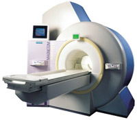
From Siemens Medical Systems;
the 3 T MAGNETOM Allegra is a dedicated MR headscanner, perfect as a research system in cognitive and neuroscience with MRS and fMRI. MAGNETOM Allegra is a full member of the MAGNETOM product family. It uses many common components, i.e. electronics, computer system, software and pulse sequence concepts.
Device Information and Specification
GRE, IR, FIR, STIR, TrueIR/FISP, FSE, FLAIR, MT, SS-FSE, MT-SE, MTC, MSE, EPI, GMR, fat/water sat./exc.
IMAGING MODES
Single, multislice, volume study, multi angle, multi oblique
178 images/sec at 256 x 256 at 100% FOV
1024 x 1024 full screen display
MAGNET WEIGHT (gantry included)
5500 kg
DIMENSION H*W*D (gantry included)
220 x 220 x 147 cm
POWER REQUIREMENTS
380/400/420/440/480 V
Passive, act.; 1st order std./2nd opt.
| | | |
• View the DATABASE results for 'MAGNETOM Allegra™' (2).
| | | | |
| | | Searchterm 'Helium' was also found in the following services: | | | | |
| | |
| |
|
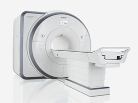
From Siemens Medical Systems;
Received FDA clearance in 2012.
The MAGNETOM Spectra is a cost-optimized high field MRI system with Tim 4G and Dot technologies. The system consumes less energy compared to other 3 Tesla scanners. The magnet-cooling helium is contained in a closed loop, which prevents the gas from escaping and reduces the need for refills. TimTX includes innovative techniques in the radio frequency excitation hardware as well as new application and processing features enabling uniform RF distribution in all body regions.
Device Information and Specification
CLINICAL APPLICATION
Whole Body
Head, spine, torso/ body coil, neurovascular, neck and multi-purpose flex coils. Peripheral vascular, breast, shoulder, knee, wrist, foot//ankle, endorectal optional.
Chemical shift imaging, single voxel spectroscopy
DIMENSION H*W*D (gantry included)
173 x 231 x 219 cm
COOLING SYSTEM
Water; single cryogen, 2 stage refrigeration
Passive, active; first order standard, second order optional
POWER REQUIREMENTS
380 / 400 / 420 / 440 / 460 / 480 V, 3-phase + ground; connection value with chiller 100 kvA /without chiller 60 kVA
| | | | | |
| | | | |
| | |
| | | |
|
| |
 | Look
Ups |
| |


