 | Info
Sheets |
| | | | | | | | | | | | | | | | | | | | | | | | |
 | Out-
side |
| | | | |
|
| | | 'Vascular Imaging with Gas' | |
Result : Searchterm 'Vascular Imaging with Gas' found in 1 term [ ] and 0 definition [ ] and 0 definition [ ], (+ 3 Boolean[ ], (+ 3 Boolean[ ] results ] results
| | 1 - 4 (of 4) Result Pages : [1] |  | | | | |  |
| Vascular Imaging with Gas |   |
| |
|
The use of gas as a contrast medium has significant potential to avoid limitations of conventional contrast agents. Gases can transit smaller vascular conduits and can be injected through smaller and less traumatic access systems than liquids. Highly soluble gases (such as CO2) can be imaged as a bolus. Blood is displaced by the gas, with the result of negative image contrast.
Because gases are compressible, standard liquid injectors
cannot be used. The design for a gasinjector should have the option for individual adaptation of blood flow rate, vessel diameter, pulse pressure, and heart rate. | | | | | | • Share the entry 'Vascular Imaging with Gas':    | | | | |  Further Reading: Further Reading: | News & More:
|
|
| |
| | | | |  |
| |
|
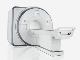
From Siemens Medical Systems;
Received FDA clearance in 2012.
The MAGNETOM Spectra is a cost-optimized high field MRI system with Tim 4G and Dot technologies. The system consumes less energy compared to other 3 Tesla scanners. The magnet-cooling helium is contained in a closed loop, which prevents the gas from escaping and reduces the need for refills. TimTX includes innovative techniques in the radio frequency excitation hardware as well as new application and processing features enabling uniform RF distribution in all body regions.
Device Information and Specification
CLINICAL APPLICATION
Whole Body
Head, spine, torso/ body coil, neuro vascular, neck and multi-purpose flex coils. Peripheral vascular, breast, shoulder, knee, wrist, foot//ankle, endorectal optional.
Chemical shift imaging, single voxel spectroscopy
DIMENSION H*W*D (gantry included)
173 x 231 x 219 cm
COOLING SYSTEM
Water; single cryogen, 2 stage refrigeration
Passive, active; first order standard, second order optional
POWER REQUIREMENTS
380 / 400 / 420 / 440 / 460 / 480 V, 3-phase + ground; connection value with chiller 100 kvA / without chiller 60 kVA
| | | | | |
| | | | | |
| |
|

Pharmacyclics is a pharmaceutical company developing novel agents to improve current therapeutic approaches to cancer, cardio vascular diseases, and inflammation.
The company's proprietary products are derived from its core technology in biometallic chemistry. These products under development are based upon small molecules, which bind metals in a unique way and are capable of capturing and focusing forms of energy used in a variety of medical applications.
The company has one product in registration with the FDA, Gadolite® Oral Suspension, a contrast agent for imaging the gastrointestinal tract; two products in clinical development, Gd-Tex ( Motexafin gadolinium), a radiation sensitizer, Lu-Tex, a photosensitizer; and several compounds in preclinical development.
MRI Contrast Agents:
Contact Information MAIL Pharmacyclics Inc.
995 East Arques Avenue
94085 Sunnyvale, California
USA | | | |
• View the DATABASE results for 'Pharmacyclics, Inc.' (2).
| | |
• View the NEWS results for 'Pharmacyclics, Inc.' (5).
| | | | | | Further Reading: | News & More:
|
|
| |
| | | | | |
| |
|
General MRI of the abdomen can consist of T1 or T2 weighted spin echo, fast spin echo ( FSE, TSE) or gradient echo sequences with fat suppression and contrast enhanced MRI techniques. The examined organs include liver, pancreas, spleen, kidneys, adrenals as well as parts of the stomach and intestine (see also gastrointestinal imaging). Respiratory compensation and breath hold imaging is mandatory for a good image quality.
T1 weighted sequences are more sensitive for lesion detection than T2 weighted sequences at 0.5 T, while higher field strengths (greater than 1.0 T), T2 weighted and spoiled gradient echo sequences are used for focal lesion detection.
Gradient echo in phase T1 breath hold can be performed as a dynamic series with the ability to visualize the blood distribution. Phases of contrast enhancement include the capillary or arterial dominant phase for demonstrating hyper vascular lesions, in liver imaging the portal venous phase demonstrates the maximum difference between the liver and hypo vascular lesions, while the equilibrium phase demonstrates interstitial disbursement for edematous and malignant tissues.
Out of phase gradient echo imaging for the abdomen is a lipid-type tissue sensitive sequence and is useful for the visualization of focal hepatic lesions, fatty liver (see also Dixon), hemochromatosis, adrenal lesions and renal masses.
The standards for abdominal MRI vary according to clinical sites based on sequence availability and MRI equipment.
Specific abdominal imaging coils and liver-specific contrast agents targeted to the healthy liver tissue improve the detection and localization of lesions.
See also Hepatobiliary Contrast Agents, Reticuloendothelial Contrast Agents, and Oral Contrast Agents.
For Ultrasound Imaging (USI) see Abdominal Ultrasound at Medical-Ultrasound-Imaging.com. | | | | | |
• View the DATABASE results for 'Abdominal Imaging' (11).
| | |
• View the NEWS results for 'Abdominal Imaging' (3).
| | | | | | Further Reading: | | Basics:
|
|
News & More:
|  |
Assessment of Female Pelvic Pathologies: A Cross-Sectional Study Among Patients Undergoing Magnetic Resonance Imaging for Pelvic Assessment at the Maternity and Children Hospital, Qassim Region, Saudi Arabia
Saturday, 7 October 2023 by www.cureus.com | | |
Higher Visceral, Subcutaneous Fat Levels Predict Brain Volume Loss in Midlife
Wednesday, 4 October 2023 by www.neurologyadvisor.com | | |
Deep Learning Helps Provide Accurate Kidney Volume Measurements
Tuesday, 27 September 2022 by www.rsna.org | | |
CT, MRI for pediatric pancreatitis interobserver agreement with INSPPIRE
Friday, 11 March 2022 by www.eurekalert.org | | |
Clinical trial: Using MRI for prostate cancer diagnosis equals or beats current standard
Thursday, 4 February 2021 by www.eurekalert.org | | |
Computer-aided detection and diagnosis for prostate cancer based on mono and multi-parametric MRI: A review - Abstract
Tuesday, 28 April 2015 by urotoday.com | | |
Nottingham scientists exploit MRI technology to assist in the treatment of IBS
Thursday, 9 January 2014 by www.news-medical.net | | |
New MR sequence helps radiologists more accurately evaluate abnormalities of the uterus and ovaries
Thursday, 23 April 2009 by www.eurekalert.org | | |
MRI identifies 'hidden' fat that puts adolescents at risk for disease
Tuesday, 27 February 2007 by www.eurekalert.org |
|
| |
| | | | |
| | | 1 - 4 (of 4) Result Pages : [1] |
| |
|
| |
 | Look
Ups |
| |


