Magnetic Resonance - Technology Information Portal Welcome to MRI Technology
Info
  Sheets

Out-
      side


 'Turbo Field Echo' 
SEARCH FOR    
 
  2 3 5 A B C D E F G H I J K L M N O P Q R S T U V W X Y Z
Result : Searchterm 'Turbo Field Echo' found in 2 terms [] and 4 definitions [], (+ 7 Boolean[] results
previous     6 - 10 (of 13)     next
Result Pages : [1]  [2]  [3]
MRI Resources 
Knee MRI - Abdominal Imaging - Mobile MRI - Artifacts - Research Labs - Coils
 
Ultrafast Gradient Echo SequenceInfoSheet: - Sequences - 
Intro, 
Overview, 
Types of, 
etc.MRI Resource Directory:
 - Sequences -
 
Ultrafast Gradient Echo Sequence Timing Diagram 
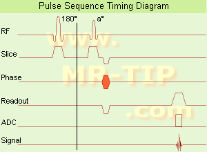
In simple ultrafast GRE imaging, TR and TE are so short, that tissues have a poor imaging signal and - more importantly - poor contrast except when contrast media enhanced (contrast enhanced angiography). Therefore, the magnetization is 'prepared' during the preparation module, most frequently by an initial 180° inversion pulse.
In the pulse sequence timing diagram, the basic ultrafast gradient echo sequence is illustrated. The 180° inversion pulse is executed one time (to the left of the vertical line), the right side represents the data collection period and is often repeated depending on the acquisition parameters.
See also Pulse Sequence Timing Diagram, there you will find a description of the components.
Ultrafast GRE sequences have a short TR,TE, a low flip angle and TR is so short that image acquisition lasts less than 1 second and typically less than 500 ms. Common TR: 3-5 msec, TE: 2 msec, and the flip angle is about 5°. Such sequences are often labeled with the prefix 'Turbo' like TurboFLASH, TurboFFE and TurboGRASS.
This allows one to center the subsequent ultrafast GRE data acquisition around the inversion time TI, where one of the tissues of interest has very little signal as its z-magnetization is passing through zero.
Unlike a standard inversion recovery (IR) sequence, all lines or a substantial segment of k-space image lines are acquired after a single inversion pulse, which can then together be considered as readout module. The readout module may use a variable flip angle approach, or the data acquisition may be divided into multiple segments (shots). The latter is useful particularly in cardiac imaging where acquiring all lines in a single segment may take too long relative to the cardiac cycle to provide adequate temporal resolution.
If multiple lines are acquired after a single pulse, the pulse sequence is a type of gradient echo echo planar imaging (EPI) pulse sequence.

See also Magnetization Prepared Rapid Gradient Echo (MPRAGE) and Turbo Field Echo (TFE).
spacer
 
• Related Searches:
    • Turbo Fast Low Angle Shot
    • Turbo Field Echo
    • TurboFLASH
    • Magnetization Prepared Rapid Gradient Echo
    • Inversion
MRI Resources 
Functional MRI - Online Books - MRI Physics - Intraoperative MRI - Used and Refurbished MRI Equipment - Implant and Prosthesis pool
 
O-SCAN™InfoSheet: - Devices -
Intro, 
Types of Magnets, 
Overview, 
etc.
 
www.fonar.com/standup.htm www.fonar.com/standup.htm O-scan is manufactured and distributed by Esaote SpA
O-scan is a compact, dedicated extremity MRI system designed for easy installation and high throughput. The complete system fits in a 9' x 10' room, doesn't need for RF or magnetic shielding and it plugs in the wall. The 0.31T permanent magnet along with dual phased array RF coils, and advanced imaging protocols provide outstanding image quality and fast 25 minute complete examinations.
Esaote North America is the exclusive distributor of the O-scan system in the USA.
Device Information and Specification
CLINICAL APPLICATION
Dedicated Extremity
CONFIGURATION
Closed
Dual phased array knee, hand, foot//ankle/elbow
PULSE SEQUENCES
SE, HSE, HFE, GE, 2dGE, ME, IR, STIR, Stir T2, GESTIR, TSE, TME, FSE STIR, FSE (T1, T2), X-Bone, Turbo 3DT1, 3D SHARC, 3D SST1, 3D SST2
IMAGING MODES
2D, 3D multi-plane, half echo, half scan, real time
TR
10 - 10,000 msec.
TE
6 - 220 msec.
SINGLE SLICE
0.1 sec.
MULTI SLICE
0.1 sec.
14 cm
2D: 2mm - 10 mm, 3D: 0.6 - 10 mm
MEASURING MATRIX
512 x 512 max.
PIXEL INTENSITY
4,096 grey levels
MAGNET TYPE
Permanent - NdFeB
MAGNET WEIGHT
2,733 lbs
POWER REQUIREMENTS
100/110/200/220/230/240
STRENGTH
20 mT/m
5 GAUSS FRINGE FIELD, radial/axial
67 cm / 75 cm
passive
spacer
MRI Resources 
Education pool - Online Books - Safety pool - Examinations - PACS - Liver Imaging
 
CHORUS 1.5T™InfoSheet: - Devices -
Intro, 
Types of Magnets, 
Overview, 
etc.MRI Resource Directory:
 - Devices -
 
www.isoltech.co.kr/english/product/default.htm 'Next generation MRI system 1.5T CHORUS developed by ISOL Technology is optimized for both clinical diagnostic imaging and for research development.
CHORUS offers the complete range of feature oriented advanced imaging techniques- for both clinical routine and research. The compact short bore magnet, the patient friendly design and the gradient technology make the innovation to new degree of perfection in magnetic resonance.'
Device Information and Specification
CLINICAL APPLICATION
Whole body
CONFIGURATION
Short bore
Head, C-spine, L-spine, TMJ, Knee, Shoulder, General purpose, Phased Array System: 4 digital receiver channels (Up to 12 channels)
Optional
SYNCHRONIZATION
ECG/peripheral: Optional/yes, respiratory gating
PULSE SEQUENCES
Spin Echo, Gradient Echo, Fast Spin Echo, Inversion Recovery (STIR, Fluid Attenuated Inversion Recovery), FLASH, FISP, PSIF, Turbo Flash ( MPRAGE ),TOF MR Angiography, Standard echo planar imaging package (SE-EPI, GE-EPI), Optional: Advanced P.A. Imaging Package (up to 4 ch.), Advanced echo planar imaging package, Single Shot and Diffusion Weighted EPI, IR/FLAIR EPI
IMAGING MODES
2D/3D, Travelling Sat, Multi-Slab 3D, MTC and TONE Pulse Sequence, Fat/Water Suppression, Presaturation (up to 6 bands), Flow Compensation using GMR pulse, Multi-Slice, Multi-Group Imaging
SINGLE/MULTI SLICE
Image reconstruction time (2562 ) : 0.02 s
FOV
40 cm
BORE DIAMETER
or W x H
58 cm diameter (patient)
MAGNET WEIGHT
4050 kg
H*W*D
233 x 206 x 160 cm
COOLING SYSTEM TYPE
Water-cooled coil and air-cooled amplifier
CRYOGEN USE
0.07 L/hr helium
STRENGTH
20 mT/m (Upto 27 mT/m)
5-GAUSS FRINGE FIELD
2.5 m / 3.8 m
Passive and active
spacer

• View the DATABASE results for 'CHORUS 1.5T™' (2).Open this link in a new window

MRI Resources 
Mobile MRI Rental - Hospitals - Anatomy - Jobs pool - Health - Veterinary MRI
 
ENCORE 0.5T™InfoSheet: - Devices -
Intro, 
Types of Magnets, 
Overview, 
etc.MRI Resource Directory:
 - Devices -
 
www.isoltech.co.kr/english/product/05t.htm 'MRI system is not an expensive equipment anymore. ENCORE developed by ISOL Technology is a low cost MRI system with the advantages like of the 1.0T MRI scanner. Developed specially for the overseas market, the ENCORE is gaining popularity in the domestic market by medium sized hospitals.
Due to the optimum RF and Gradient application technology. ENCORE enables to obtain high resolution imaging and 2D/3D Angio images which was only possible in high field MR systems.'
- Less consumption of the helium gas due to the ultra-lightweight magnet specially designed and manufactured for ISOL. - Cost efficiency MR system due to air cooling type (equivalent to permanent magnetic). - Patient processing speed of less than 20 minutes.'
Device Information and Specification
CLINICAL APPLICATION
Whole body
CONFIGURATION
Short bore compact
Head, C-spine, L-spine, TMJ, Knee, Shoulder, General purpose, Phased Array System: 4 digital receiver channels (Up to 12 channels)
SYNCHRONIZATION
ECG/peripheral: Optional/yes, respiratory gating
SINGLE/MULTI SLICE
Image reconstruction time (2562 ) : 0.02 s
FOV
40 cm
BORE DIAMETER
or W x H
58 cm diameter
MAGNET WEIGHT
3200 kg
H*W*D
200 x 168 x 187 cm
COOLING SYSTEM TYPE
Air-cooled coil and amplifier
CRYOGEN USE
0.05 L/hr helium
STRENGTH
15 mT/m
5-GAUSS FRINGE FIELD
2.3 m / 3.1 m
Passive and active
spacer

• View the DATABASE results for 'ENCORE 0.5T™' (2).Open this link in a new window

MRI Resources 
Homepages - Corporations - Resources - Raman Spectroscopy - Cardiovascular Imaging - Education
 
Lung ImagingMRI Resource Directory:
 - Lung Imaging -
 
Lung imaging is furthermore a challenge in MRI because of the predominance of air within the lungs and associated susceptibility issues as well as low signal to noise of the inflated lung parenchyma. Cardiac and respiratory triggered or breath hold sequences allow diagnostic imaging, however a comparable image quality with computed tomography is still difficult to achieve.
Assumptions for lung MRI:
•
Low signal to noise ratio of the inherently low lung proton density.
•
Cardiac and respiratory motion artifacts.
•
Magnetic susceptibility effects of large magnetic field gradients.
•
Very short transverse relaxation times and significant diffusion yielding short T2 (30-70 msec), short T2* (1-3 msec), and additional long T1 relaxation times (1300-1500 msec).
•
The extreme short T2 values are responsible for a fast signal decay during a single shot readout, resulting in blurring.

The current trends in MRI are the use of new imaging technologies and increasingly powerful magnetic fields. Among these technologies are parallel imaging techniques as well as ventilation agents like hyperpolarized helium for the use as an inert inhalational contrast agent to study lung ventilation properties. With hyperpolarized gases clear images of the lungs can be obtained without using a large magnetic field (see also back projection imaging). Single shot sequences (e.g. TSE or Half Fourier Acquisition Single Shot Turbo Spin Echo HASTE) used in lung MR imaging benefits from parallel imaging techniques due to reduced relaxation time effects during the echo train and therefore reduced image blurring as well as reduced motion artifacts.
In the future, more effective contrast agents may provide an alternative solution to the need for high field MRI. Dynamic contrast enhanced MRI perfusion has demonstrated a potential in the diagnosis of pulmonary embolism or to characterize lung cancer and mediastinal tumors. 3D contrast enhanced magnetic resonance angiography of the thoracic vessel.

See also the related poll result: 'MRI will have replaced 50% of x-ray exams by'
 
Images, Movies, Sliders:
 Anatomic Imaging of the Lungs  Open this link in a new window
      

Courtesy of  Robert R. Edelman
 Normal Lung Gd Perfusion MRI  Open this link in a new window
      

Courtesy of  Robert R. Edelman

 MRI Thorax Basal Plane  Open this link in a new window
 
Radiology-tip.comradLung Scintigraphy
spacer

• View the DATABASE results for 'Lung Imaging' (7).Open this link in a new window


• View the NEWS results for 'Lung Imaging' (3).Open this link in a new window.
 
Further Reading:
  Basics:
A safer approach for diagnostic medical imaging
Monday, 29 September 2014   by www.eurekalert.org    
Parallel Lung Imaging(.pdf)
  News & More:
Chest MRI a viable alternative to chest CT in COVID-19 pneumonia follow-up
Monday, 21 September 2020   by www.healthimaging.com    
CT Imaging Features of 2019 Novel Corona virus (2019-nCoV)
Tuesday, 4 February 2020   by pubs.rsna.org    
Polarean Imaging Phase III Trial Results Point to Potential Improvements in Lung Imaging
Wednesday, 29 January 2020   by www.diagnosticimaging.com    
Low Power MRI Helps Image Lungs, Brings Costs Down
Thursday, 10 October 2019   by www.medgadget.com    
Chest MRI Using Multivane-XD, a Novel T2-Weighted Free Breathing MR Sequence
Thursday, 11 July 2019   by www.sciencedirect.co    
Researchers Review Importance of Non-Invasive Imaging in Diagnosis and Management of PAH
Wednesday, 11 March 2015   by lungdiseasenews.com    
New MRI Approach Reveals Bronchiectasis' Key Features Within the Lung
Thursday, 13 November 2014   by lungdiseasenews.com    
MRI techniques improve pulmonary embolism detection
Monday, 19 March 2012   by medicalxpress.com    
  News & More:
Partnership with VIDA to streamline adoption of advanced MRI of the lungs
Monday, 11 September 2023   by www.itnonline.com    
MRI Resources 
PACS - Societies - Knee MRI - Pediatric and Fetal MRI - Open Directory Project - Bioinformatics
 
previous      6 - 10 (of 13)     next
Result Pages : [1]  [2]  [3]
 Random Page
 
Share This Page
FacebookTwitterLinkedIn

MR-TIP    
Community   
User
Pass
Forgot your UserID/Password ?    


How AI will impact MRI :
only diagnostics 
saving time 
reducing cost 
makes planning obsolete 
reduce human knowledge 
not at all 

Look
      Ups


MR-TIP.com uses cookies! By browsing MR-TIP.com, you agree to our use of cookies.

Magnetic Resonance - Technology Information Portal
Member of SoftWays' Medical Imaging Group - MR-TIP • Radiology-TIP • Medical-Ultrasound-Imaging • 
Copyright © 2003 - 2024 SoftWays. All rights reserved. [ 3 May 2024]
Terms of Use | Privacy Policy | Advertising
 [last update: 2024-02-26 03:41:00]