 | Info
Sheets |
| | | | | | | | | | | | | | | | | | | | | | | | |
 | Out-
side |
| | | | |
|
| | | | |
Result : Searchterm 'Real Time' found in 2 terms [ ] and 5 definitions [ ] and 5 definitions [ ], (+ 13 Boolean[ ], (+ 13 Boolean[ ] results ] results
| | previous 6 - 10 (of 20) nextResult Pages : [1] [2] [3 4] |  | |  | Searchterm 'Real Time' was also found in the following services: | | | | |
| |  |
| |
|
(PWI - Perfusion Weighted Imaging) Perfusion MRI techniques (e.g. PRESTO - Principles of Echo Shifting using a Train of Observations) are sensitive to microscopic levels of blood flow. Contrast enhanced relative cerebral blood volume (rCBV) is the most used perfusion imaging.
Both, the ready availability and the T2* susceptibility effects of gadolinium, rather than the T1 shortening effects make gadolinium a suitable agent for use in perfusion imaging. Susceptibility here refers to the loss of MR signal, most marked on T2* ( gradient echo)-weighted and T2 ( spin echo)-weighted sequences, caused by the magnetic field-distorting effects of paramagnetic substances.
T2* perfusion uses dynamic sequences based on multi or single shot techniques. The T2* ( T2) MRI signal drop within or across a brain region is caused by spin dephasing during the rapid passage of contrast agent through the capillary bed. The signal decrease is used to compute the relative perfusion to that region. The bolus through the tissue is only a few seconds, high temporal resolution imaging is required to obtain sequential images during the wash in and wash out of the contrast material and therefore, resolve the first pass of the tracer. Due to the high temporal resolution, processing and calculation of hemodynamic maps are available (including mean transit time (MTT), time to peak (TTP), time of arrival (T0), negative integral (N1) and index.
An important neuroradiological indication for MRI is the evaluation of incipient or acute stroke via perfusion and diffusion imaging. Diffusion imaging can demonstrate the central effect of a stroke on the brain, whereas perfusion imaging visualizes the larger 'second ring' delineating blood flow and blood volume. Qualitative and in some instances quantitative (e.g. quantitative imaging of perfusion using a single subtraction) maps of regional organ perfusion can thus be obtained.
Echo planar and potentially echo volume techniques together with appropriate computing power offer real time images of dynamic variations in water characteristics reflecting perfusion, diffusion, oxygenation (see also Oxygen Mapping) and flow.
Another type of perfusion MR imaging allows the evaluation of myocardial ischemia during pharmacologic stress. After e.g., adenosine infusion, multiple short axis views (see cardiac axes) of the heart are obtained during the administration of gadolinium contrast. Ischemic areas show up as areas of delayed and diminished enhancement. The MRI stress perfusion has been shown to be more accurate than nuclear SPECT exams. Myocardial late enhancement and stress perfusion imaging can also be performed during the same cardiac MRI examination. | | | | | | | | | | | | | | | | | |  Further Reading: Further Reading: | | Basics:
|
|
News & More:
| |
| |
| | | Searchterm 'Real Time' was also found in the following service: | | | | |
| | |
| |
|

From GE Healthcare;
The Signa HDx MRI system is GE's leading edge whole body magnetic resonance scanner designed to support high resolution, high signal to noise ratio, and short scan times.
Signa HDx 3.0T offers new technologies like ultra-fast image reconstruction through the new XVRE recon engine, advancements in parallel imaging algorithms and the broadest range of premium applications. The HD applications, PROPELLER (high-quality brain imaging extremely resistant to motion artifacts), TRICKS (contrast-enhanced angiographic vascular lower leg imaging), VIBRANT (for breast MRI), LAVA (high resolution liver imaging with shorter breath holds and better organ coverage) and MR Echo (high-definition cardiac images in real time) offer unique capabilities.
Device Information and Specification CLINICAL APPLICATION Whole body
CONFIGURATION Compact short bore SE, IR, 2D/3D GRE, RF-spoiled GRE, 2DFGRE, 2DFSPGR, 3DFGRE, 3DFSPGR, 3DTOFGRE, 3DFSPGR, 2DFSE, 2DFSE-XL, 2DFSE-IR, T1-FLAIR, SSFSE, EPI, DW-EPI, BRAVO, Angiography: 2D/3D TOF, 2D/3D phase contrast vascular IMAGING MODES Single, multislice, volume study, fast scan, multi slab, cine, localizer H*W*D 240 x 2216,6 x 201,6 cm POWER REQUIREMENTS 480 or 380/415, 3 phase ||
COOLING SYSTEM TYPE Closed-loop water-cooled grad. | | | | | |
| | | | |  |
| |
|

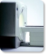
Manufactured by Esaote S.p.A.;
a low field open MRI scanner with permanent magnet for orthopedic use. The outstanding feature of this MRI system is a patient friendly design with 24 cm diameter, which allows the imaging of extremities and small body parts like shoulder MRI. The power consumption is around 1.3 kW and the needed minimum floor space is an area of 16 sq m.
At RSNA 2006 Hologic Inc. introduced a new dedicated extremity MRI scanner, the Opera. Manufactured by Esaote is the Opera a redesign of Esaote's 0.2 Tesla E-Scan XQ platform, which now enables complete imaging of all extremities, including hip and shoulder applications. ' Real- time positioning' reportedly speeds patient setup and reduces exam times.
Esaote North America and Hologic Inc are the U.S. distributors of this MRI device.
Device Information and Specification CLINICAL APPLICATION Dedicated extremity
SE, GE, IR, STIR, FSE, 3D CE, GE-STIR, 3D GE, ME, TME, HSE IMAGING MODES Single, multislice, volume study, fast scan, multi slab2D: 2 mm - 10 mm;
3D: 0.6 mm - 10 mm 4096 gray lvls, 256 lvls in 3D POWER REQUIREMENTS 2,0 kW; 110/220 V single phase | | | |
• View the DATABASE results for 'Opera (E-SCAN™ XQ)' (2).
| | | | | | Further Reading: | News & More:
|
|
| |
| | | Searchterm 'Real Time' was also found in the following services: | | | | |
| | |
| |
|

(Signa VH/i 3.0T)
With GE Healthcare
leading-edge technology in ultra-high-field imaging. The 3 T VH/i provides a platform for advanced applications in radiology, cardiology, psychology and psychiatry. Real- time image processing lets you acquire multislice whole brain images and map brain functions for research or surgical planning. And the 3 T Signa VH/i is flexible enough to provide clinicians with high performance they require. It can provide not only outstanding features in brain scanning and neuro-system research, but also a wide range of use in scanning breasts, extremities, the spine and the cardiovascular systems.
Device Information and Specification CLINICAL APPLICATION Whole body
T/R quadrature head, T/R quadrature body, T/R phased array extremity (opt) SE, IR, 2D/3D GRE, FGRE, RF-spoiled GRE, FSE, Angiography: 2D/3D TOF, 2D/3D phase contrast vascular IMAGING MODES Single, multislice, volume study, fast scan, multi slab, cine, localizer 100 Images/sec with Reflex100 MULTISLICE 100 Images/sec with Reflex100 2D 0.5-100mm in 0.1mm incremental 128x512 steps 32 phase encode H*W*D 260cm x 238cm x 265cm POWER REQUIREMENTS 480 or 380/415, 3 phase ||
COOLING SYSTEM TYPE Closed-loop water-cooled grad. Less than 0.14 L/hr liquid He | | | |
• View the DATABASE results for 'Signa 3.0T™' (2).
| | | | |
| | | Searchterm 'Real Time' was also found in the following service: | | | | |
| | |
| |
|
From GE Healthcare;
GE's Signa Contour/i system uses the innovations like K4 technology and real- time interactive imaging.
This compact magnet with wide-flare gantry obtains high patient comfort with low costs.
Device Information and Specification CLINICAL APPLICATION Whole body Head and body coil standard; all other coils optional; open architecture makes system compatible with a wide selection of coils Standard: SE, IR, 2D/3D GRE and SPGR, Angiography;; 2D/3D TOF, 2D/3D Phase Contrast;; 2D/3D FSE, 2D/3D FGRE and FSPGR, SSFP, FLAIR, optional: EPI, 2D/3D Fiesta, FGRET, Spiral2D 0.8 mm to 20 mm; 3D 0.1 mm to 5 mm 128x512 steps 32 phase encode POWER REQUIREMENTS 480 or 380/415 V STRENGTH SmartSpeed 23 mT/m, HiSpeed Plus 33 mT/m | | | | | |
| | | | |
| | | |
|
| |
 | Look
Ups |
| |


