Magnetic Resonance - Technology Information Portal Welcome to MRI Technology
Info
  Sheets

Out-
      side


 'Passive Shimming' 
SEARCH FOR    
 
  2 3 5 A B C D E F G H I J K L M N O P Q R S T U V W X Y Z
Result : Searchterm 'Passive Shimming' found in 1 term [] and 3 definitions [], (+ 17 Boolean[] results
previous     16 - 20 (of 21)     next
Result Pages : [1]  [2 3 4 5]
MRI Resources 
Musculoskeletal and Joint MRI - Health - Contrast Enhanced MRI - Spine MRI - Coils - Claustrophobia
 
Virgoâ„¢InfoSheet: - Devices -
Intro, 
Types of Magnets, 
Overview, 
etc.MRI Resource Directory:
 - Devices -
 
www.canamglobal.com/mri/virgomri.html From Millennium Technology Inc. 
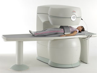
This open C-shaped MRI system eases patient comfort and technologist maneuverability. This low cost scanner is build for a wide range of applications. The Virgo™ patient table is detachable and moves on easy rolling castors. Able to accommodate patient weights up to 160 kg, the tabletop has a range of motion of 30 cm in the lateral direction and 90cm in the longitudinal direction. Images generated with this scanner can only be viewed (without data loss) on Millennium's proprietary viewing software.
Device Information and Specification
CLINICAL APPLICATION
Whole body
CONFIGURATION
C shaped
SYNCHRONIZATION
Standard cardiac gating, ECG/peripheral, respiratory gating
PULSE SEQUENCES
2D Spin Echo (single and multi-echo), 2D Inversion Recovery, 2D Sequential and 3D Volume Gradient Echo, 2D and 3D Spoiled Gradient Echo
IMAGING MODES
Localizer, single slice, multislice, volume, fast, POMP, multi slab, cine, slice and frequency zip, extended dynamic range, tailored RF
TR
steps of 1 msec
TE
steps of 1 msec
SINGLE/MULTI SLICE
Simultaneous scan and reconstruction;; 100 images/second reconstruction
400 mm
2D : 2 mm; 3D : 0.5 mm
MEASURING MATRIX
512x512
PIXEL INTENSITY
256 gray levels
MAGNET TYPE
Permanent
BORE DIAMETER
or Vertical Gap
44 cm
STRENGTH
15 mT/m
5-GAUSS FRINGE FIELD
3 m/3 m
Passive
spacer
MRI Resources 
MRI Training Courses - Musculoskeletal and Joint MRI - NMR - Patient Information - MRI Technician and Technologist Schools - Most Wanted
 
S-SCANInfoSheet: - Devices -
Intro, 
Types of Magnets, 
Overview, 
etc.
 
www.esaote.com/products/MRI/sScan/products1.htm From Esaote S.p.A.; Esaote introduced the S-SCAN at RSNA in November 2007. The S-SCAN is a dedicated joint and spine MR scanner derived from the company's earlier G-SCAN system. Unlike the G-SCAN, neither the patient table nor the magnet can rotate from horizontal to vertical position. The patient table can only moved manually. Improved electronics, new coils for lumbar and cervical spine, new pulse sequences, a modified version of the magnet poles and gradient coils are used with a new software release in the S-SCAN.
Esaote North America is the exclusive U.S. distributor of this MRI device.
Device Information and Specification
CLINICAL APPLICATION
CONFIGURATION
4-channel phased array spine coil, extremity, shoulder, flex coil, knee dual phased array, ankle//foot dual phased array, hand//wrist dual phased array
PULSE SEQUENCES
SE, GE, IR, STIR, TSE, 3D CE, GE-STIR, 3D GE, ME, TME, HSE
IMAGING MODES
Single, multislice, volume study, fast scan, multi slab, cine
25 cm
MEASURING MATRIX
256 x 256 maximum
MAGNET TYPE
Permanent
33 cm H, open
POWER REQUIREMENTS
3 kW; 110/220 V single phase
STRENGTH
25 mT/m
5-GAUSS FRINGE FIELD, radial/axial
180 cm
Passive
spacer

• View the DATABASE results for 'S-SCAN' (3).Open this link in a new window


• View the NEWS results for 'S-SCAN' (1).Open this link in a new window.
 
Further Reading:
  News & More:
Wichita medical facility gets first Hologic S-scan MRI in the United States
Friday, 19 October 2007   by wichita.bizjournals.com    
MRI Resources 
Raman Spectroscopy - IR - Cochlear Implant - Most Wanted - Homepages - Health
 
G-SCANInfoSheet: - Devices -
Intro, 
Types of Magnets, 
Overview, 
etc.MRI Resource Directory:
 - Devices -
 
www.esaote.de/04_kernspin/gscan/gscan.htm From Esaote S.p.A.; Esaote introduced the new G-SCAN at the RSNA in Dec. 2004. The G-SCAN covers almost all musculoskeletal applications including the spine. The tilting gantry is designed for scanning in weight-bearing positions. This unique MRI scanner is developed in line with the Esaote philosophy of creating high quality MRI systems that are easy to install and that have a low breakeven point.
Device Information and Specification
CLINICAL APPLICATION
CONFIGURATION
Spine, extremity, shoulder, flex coil, knee dual phased array, ankle//foot dual phased array, hand//wrist dual phased array
PULSE SEQUENCES
SE, GE, IR, STIR, TSE, 3D CE, GE-STIR, 3D GE, ME, TME, HSE
IMAGING MODES
Single, multislice, volume study, fast scan, multi slab, cine
FOV
100 up to 350 mm, 25 mm displayed
512 x 512
MEASURING MATRIX
256 x 256 maximum
MAGNET TYPE
Permanent
BORE DIAMETER
or W x H
33 cm H, open
POWER REQUIREMENTS
100/110/200/220/230/240 V
STRENGTH
25 mT/m
5-GAUSS FRINGE FIELD
180 cm
Passive
spacer

• View the DATABASE results for 'G-SCAN' (3).Open this link in a new window

MRI Resources 
RIS - Spectroscopy - Implant and Prosthesis pool - Jobs - Mobile MRI Rental - Quality Advice
 
FORTE 3.0Tâ„¢InfoSheet: - Devices -
Intro, 
Types of Magnets, 
Overview, 
etc.MRI Resource Directory:
 - Devices -
 
www.isoltech.co.kr/english/product/30t.htm From ISOL Technology
'Ultra high field MR system, it's right close to you. FORTE 3.0T is the new standard for the future ultra high field MR system. If you are pushing the limits of your existing clinical MR scanner, the FORTE will surely take you to the next level of diagnostic imaging. FORTE is the core leader of the medical technology in the 21st century. Proving effects of fMRI that cannot be measured with MRI less than 2.0T.'
Device Information and Specification
CLINICAL APPLICATION
Whole body
CONFIGURATION
Short bore compact
SYNCHRONIZATION
ECG/peripheral: Optional/yes, external trigger, respiratory gating
PULSE SEQUENCES
Spin echo, Gradient echo, Fast spin echo, Inversion recovery, 2D/3D Fast gradient echo sequences FLAIR/STIR, 2D/3D TOF
IMAGING MODES
2D/3D, T1, T2 and Diffusion//Perfusion imaging, MR Angiography package, Advanced EPI package, Multi-nuclei MR Spectroscopy package
FOV
40 cm
128 x 128, 256 x 256, 512 x 512, 1024 x 1024
BORE DIAMETER
or W x H
61 cm without body coil
MAGNET WEIGHT
12000 kg
H*W*D
260 x 220 x 235 cm
COOLING SYSTEM TYPE
Water-cooled coil and air-cooled amplifier
CRYOGEN USE
0.15 L/hr helium
STRENGTH
38 mT/m
5-GAUSS FRINGE FIELD
3.3 m / 5.2 m
Passive and active
spacer

• View the DATABASE results for 'FORTE 3.0T™' (2).Open this link in a new window

MRI Resources 
Spectroscopy - Guidance - Online Books - MRI Technician and Technologist Career - Hospitals - Functional MRI
 
Panorama 0.6TPanorama 0.2InfoSheet: - Devices -
Intro, 
Types of Magnets, 
Overview, 
etc.MRI Resource Directory:
 - Devices -
 
www.medical.philips.com/main/products/mri/products/panoramafamily/panorama0.23t_rt/features/ From Philips Medical Systems;
Panorama 0.6 T is the Philips Mid-Field Open MRI system. It is the most open MR scanner in the market, optimized for patient comfort and faster reconstruction time.
Device Information and Specification
CLINICAL APPLICATION
Whole body
CONFIGURATION
Open MRI/C-arm
Head, head-neck, extremity, neck, body/spine M-XL, shoulder, bilateral breast, wrist, TMJ, flex XS-S-M-L-XL-XXL
SYNCHRONIZATION
ECG/peripheral: Optional/yes, respiratory gating
PULSE SEQUENCES
SE, FE, IR, STIR, FFE, DEFFE, DESE, TSE, DETSE, Single shot SE, DRIVE, Balanced FFE, MRCP, Fluid Attenuated Inversion Recovery, Turbo FLAIR, IR-TSE, T1-STIR TSE, T2-STIR TSE, Diffusion Imaging, 3D SE, 3D FFE, Contrast Perfusion Analysis, MTC;; Angiography: CE-ANGIO, MRA 2D, 3D TOF
IMAGING MODES
Single, multislice, volume study, dynamic, SIMEX, multi chunk 3D, multiple stacks
TR
Min. 4.6 msec
TE
Min. 2.3 msec
SINGLE/MULTI SLICE
50 slices/sec
0.4 cm - 42 cm
1280 X 1024
MEASURING MATRIX
Up to 512 x 512
PIXEL INTENSITY
256 gray scale
MAGNET TYPE
Superconducting / iron core
Open x 47 cm x infinite (side-first patient entry)
MAGNET WEIGHT
38000 kg
H*W*D
254 x 244 x 325 cm
POWER REQUIREMENTS
400/480 V
COOLING SYSTEM TYPE
Liquid helium//air cool
0.00 L/hr helium
STRENGTH
20 mT/m
5-GAUSS FRINGE FIELD
2.4 m / 2.5 m
Passive/active
spacer

• View the DATABASE results for 'Panorama 0.6T™' (2).Open this link in a new window

MRI Resources 
Distributors - Databases - Implant and Prosthesis - Knee MRI - Online Books - Bioinformatics
 
previous      16 - 20 (of 21)     next
Result Pages : [1]  [2 3 4 5]
 Random Page
 
Share This Page
FacebookTwitterLinkedIn

MR-TIP    
Community   
User
Pass
Forgot your UserID/Password ?    


Personalized protocols (age, gender, body habitus, etc.) lead to :
more automated planning 
improved patient comfort 
better diagnostics 
optimized image quality 
nothing 

Look
      Ups


MR-TIP.com uses cookies! By browsing MR-TIP.com, you agree to our use of cookies.

Magnetic Resonance - Technology Information Portal
Member of SoftWays' Medical Imaging Group - MR-TIP • Radiology-TIP • Medical-Ultrasound-Imaging • 
Copyright © 2003 - 2024 SoftWays. All rights reserved. [ 29 April 2024]
Terms of Use | Privacy Policy | Advertising
 [last update: 2024-02-26 03:41:00]