Magnetic Resonance - Technology Information Portal Welcome to MRI Technology
Info
  Sheets

Out-
      side


 'Gradient Pulse' 
SEARCH FOR    
 
  2 3 5 A B C D E F G H I J K L M N O P Q R S T U V W X Y Z
Result : Searchterm 'Gradient Pulse' found in 3 terms [] and 31 definitions []
previous     6 - 10 (of 34)     next
Result Pages : [1]  [2 3 4 5 6 7]
Searchterm 'Gradient Pulse' was also found in the following services: 
spacer
News  (3)  Resources  (1)  Forum  (1)  
 
Incoherent Gradient Echo (RF Spoiled)InfoSheet: - Sequences - 
Intro, 
Overview, 
Types of, 
etc.
 
A gradient echo is generated by using a pair of bipolar gradient pulses. The gradient field is negatively pulsed, causing the spins of the xy-magnetization to dephase. A second gradient pulse is applied with the opposite polarity. During the pulsing, the spins that dephased begin to rephase and generate a gradient echo.
Spoiling can be accomplished by RF or a gradient. The incoherent RF spoiled type of a gradient echo sequence use a continuous shifting of the RF pulse to spoil the residual transverse magnetization. The phase of the RF excitation and receiver channel are varied pseudo randomly with each excitation cycle to prevent the xy magnetization from achieving steady state. T2* does not dominate image contrast, so T1 and PD weighting is practical. This method is effective and can be used to achieve a shorter TR, due to a lack of additional gradients. Spoiling eliminates the effect of the remaining xy-magnetization and leads to steady state longitudinal magnetization. These sequences can be used for breath hold, dynamic imaging and in cine and volume acquisitions.
spacer
 
• Related Searches:
    • Ernst Angle
    • Bipolar Gradient Pulse
    • Spoiled Gradient Echo Sequence
    • Radio Frequency Pulse
    • Gradient Recalled Acquisition in Steady State
MRI Resources 
RIS - Used and Refurbished MRI Equipment - Service and Support - Societies - Nerve Stimulator - Pacemaker
 
Phase Contrast SequenceMRI Resource Directory:
 - Sequences -
 
(PC) Phase contrast sequences are the basis of MRA techniques utilizing the change in the phase shifts of the flowing protons in the region of interest to create an image. Spins that are moving along the direction of a magnetic field gradient receive a phase shift proportional to their velocity.
In a phase contrast sequence two data sets with a different amount of flow sensitivity are acquired. This is usually accomplished by applying gradient pairs, which sequentially dephase and then rephase spins during the sequence. Both 2D and 3D acquisition techniques can be applied with phase contrast MRA.
The first data set is acquired with a flow compensated sequence, i. e. without flow sensitivity. The second data set is acquired with a flow sensitive sequence. The amount of flow sensitivity is controlled by the strength of the bipolar gradient pulse pair, which is incorporated into the sequence. Stationary tissue undergoes no effective phase change after the application of the two gradients. Caused by the different spatial localization of flowing blood to stationary tissue, it experiences a different size of the second bipolar gradient compared to the first. The result is a phase shift.
The raw data from the two data sets are subtracted. By comparing the phase of signals from each location in the two sequences the exact amount of motion induced phase change can be determined to have a map where pixel brightness is proportional to spatial velocity.
Phase contrast images represent the signal intensity of the velocity of spins at each point within the field of view. Regions that are stationary remain black while moving regions are represented as grey to white.
The phase shift is proportional to the spin's velocity, and this allows the quantitative assessment of flow velocities. The difference MRI signal has a maximum value for opposite directions. This velocity is typically referred to as venc, and depends on the pulse amplitude and distance between the gradient pulse pair. For velocities larger than venc the difference signal is decreased constantly until it gets zero. Therefore, in a phase contrast angiography it is important to correctly set the venc of the sequence to the maximum flow velocity which is expected during the measurement. High venc factors of the PC angiogram (more than 40 cm/sec) will selectively image the arteries (PCA - arteriography), whereas a venc factor of 20 cm/sec will perform the veins and sinuses (PCV or MRV - venography).

See also Flow Quantification, Contrast Enhanced MR Venography, Time of Flight Angiography, Time Resolved Imaging of Contrast Kinetics.
 
Images, Movies, Sliders:
 PCA-MRA 3D Brain Venography Colored MIP  Open this link in a new window


spacer

• View the DATABASE results for 'Phase Contrast Sequence' (5).Open this link in a new window

 
Further Reading:
  Basics:
MR–ANGIOGRAPHY(.pdf)
MRI Resources 
MRI Training Courses - Absorption and Emission - Abdominal Imaging - Journals - Veterinary MRI - MR Guided Interventions
 
Pulse Sequence Timing DiagramInfoSheet: - Sequences - 
Intro, 
Overview, 
Types of, 
etc.
 
Spin Echo Timing Diagram 
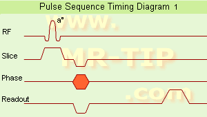
The schematic figures of a
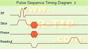
 pulse sequence timing diagram illustrate the steps of basic hardware activity that are incorporated into a pulse sequence. Time during sequence execution is indicated along the horizontal axes. Each line belongs to a different hardware component. One line is needed for the radio frequency transmitter and also one for each gradient (Gs = slice selection gradient x, Gf = phase encoding gradient y, Gf = frequency encoding gradient z, also called readout gradient).
In picture 1, a timing diagram for a 2D pulse sequence is shown.
Slice selection and signal detection are repeated in duration, relative timing and amplitude, each time the sequence is repeated. A single phase encoding component is present each time the sequence is executed.
Additional lines are added for ADC (Analog to Digital Converter) and sampling. A gradient pulse is shown as a deviation above or below the horizontal line. Simultaneous component activities such as the RF pulse and slice selection gradient are indicated as a non-zero deviation from both lines at the same horizontal position. Simple deviations from zero show constant amplitude gradient pulse. Gradient amplitudes that change during the measurement, e.g. phase encoding are represented as hatched regions.

Spin Echo Timing Diagram The second picture shows a timing diagram for a 3D pulse sequence.
Volume excitation and signal detection are repeated in duration, relative timing and amplitude, each time the sequence is repeated. Two phase encoding components are present, one in the phase encoding direction and the other in slice selection direction (irrespectively incremented in amplitude) in each time the sequence is executed. A description of the comparison of hardware activity between different pulse sequences.
spacer

• View the DATABASE results for 'Pulse Sequence Timing Diagram' (7).Open this link in a new window

Searchterm 'Gradient Pulse' was also found in the following services: 
spacer
News  (3)  Resources  (1)  Forum  (1)  
 
Saturation
 
In MR, saturation is a nonequilibrium state with no net magnetization. The same amount of nuclear spins is aligned against and with the magnetic field. Saturation methods like FatSat, SPIR etc., work with a frequency selective saturation pulse for a specific chemical shift applied before the actual sequence starts. This saturation pulse adjusts the magnetization from tissue components to zero. The hydrogen nuclei of fat and water resonate at different frequencies, which makes it possible to excite just the fat with repeatedly applying RF pulses at the Larmor frequency with interpulse times compared to T1. The resulting signal is then destroyed with a gradient pulse (Spoiler Gradient Pulse). Fat is the chemical compound to be saturated at a fat saturation sequence. When the actual sequence follows, (e.g., a spin echo sequence) the unwanted suppressed component will not resonate.

See also Saturation Recovery.
spacer

• View the DATABASE results for 'Saturation' (54).Open this link in a new window

MRI Resources 
Pregnancy - Journals - Services and Supplies - Examinations - Databases - MRCP
 
Acoustic Noise
 
Vibrations of the gradient coil support structure create sound waves. These are caused by the interactions of the magnetic field created by pulses of the current through the gradient coil with the main magnetic field in a manner similar to a loudspeaker coil. The sounds made by the scanner vary in volume and tone with the type of procedure being performed.
Sound pressure is reported on a logarithmic scale called sound-pressure level, expressed in decibel (dB) referenced to the weakest audible 1 000 Hz sound pressure of 2 * 10-5 pascal (20 micropascal). Sound level meters contain filters that simulate the ear's frequency response. The most commonly used filter provides what is called 'A' weighting, with the letter 'A' appended to the dB units, i.e. dBA.
MRI system noise levels increase with field strength. Disposable earplugs and/or headphones for the patient are recommended in high-field systems. Noise-canceling systems and special earphones are available, and active acoustic control systems were developed, e.g. softtone, pianissimo. A sequence with low noise gradient pulses is also called 'whisper sequence'.

See also Phon and Decibel.
spacer

• View the DATABASE results for 'Acoustic Noise' (9).Open this link in a new window

 
Further Reading:
  Basics:
MRI Noise in Utero Not Harmful for Baby's Ears
Tuesday, 28 September 2010   by www.medgadget.com    
  News & More:
Noise from Magnetic Resonance Imaging Can Have Short-Term Impact on Hearing
Thursday, 22 February 2018   by www.diagnosticimaging.com    
Echo Planar Imaging at 4 Tesla With Minimum Acoustic Noise(.pdf)
   by www.bnl.gov    
TOSHIBA SHOWCASES PATIENT-FRIENDLY 3T MR SYSTEM
Sunday, 29 November 2009   by medical.toshiba.com    
MRI Resources 
Bioinformatics - Universities - Calculation - Pathology - Colonography - Sequences
 
previous      6 - 10 (of 34)     next
Result Pages : [1]  [2 3 4 5 6 7]
 Random Page
 
Share This Page
FacebookTwitterLinkedIn

MR-TIP    
Community   
User
Pass
Forgot your UserID/Password ?    


New acceleration techniques will :
reduce scan times 
cause artifacts 
increase expenses 
be useful if you have a lot of experience 
doesn't do much 
never heard of 

Look
      Ups


MR-TIP.com uses cookies! By browsing MR-TIP.com, you agree to our use of cookies.

Magnetic Resonance - Technology Information Portal
Member of SoftWays' Medical Imaging Group - MR-TIP • Radiology-TIP • Medical-Ultrasound-Imaging • 
Copyright © 2003 - 2024 SoftWays. All rights reserved. [ 29 April 2024]
Terms of Use | Privacy Policy | Advertising
 [last update: 2024-02-26 03:41:00]