Magnetic Resonance - Technology Information Portal Welcome to MRI Technology
Info
  Sheets

Out-
      side


 'Gradient Magnetic Field' 
SEARCH FOR    
 
  2 3 5 A B C D E F G H I J K L M N O P Q R S T U V W X Y Z
Result : Searchterm 'Gradient Magnetic Field' found in 2 terms [] and 7 definitions [], (+ 18 Boolean[] results
previous     16 - 20 (of 27)     next
Result Pages : [1]  [2]  [3 4 5 6]
Searchterm 'Gradient Magnetic Field' was also found in the following services: 
spacer
News  (1)  Resources  (3)  
 
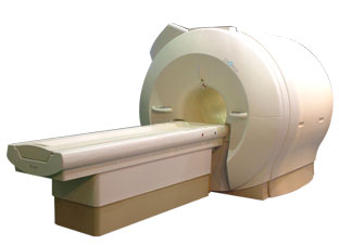
Signa Infinity 1.0T™InfoSheet: - Devices -
Intro, 
Types of Magnets, 
Overview, 
etc.MRI Resource Directory:
 - Devices -
 
www.gehealthcare.com/usen/mr/index.html From GE Healthcare;
the Signa Infinity Magnetic Resonance system is a short bore, high performance, whole-body imaging system operating at 1.0 Tesla. The system can image in any orthogonal or oblique plane (including single and double axis oblique), using a wide variety of pulse sequences.
Device Information and Specification
CLINICAL APPLICATION
Whole body
CONFIGURATION
Short bore
Head and body coil standard; all other coils optional; open architecture makes system compatible with a wide selection of coils
SYNCHRONIZATION
ECG/peripheral, respiratory gating
PULSE SEQUENCES
Standard: SE, IR, 2D/3D GRE and SPGR, Angiography;; 2D/3D TOF, 2D/3D Phase Contrast;; 2D/3D FSE, 2D/3D FGRE and FSPGR, SSFP, FLAIR, optional: EPI, 2D/3D Fiesta, FGRET, Spiral
IMAGING MODES
Localizer, single slice, multislice, volume, fast, POMP, multi slab, cine
TR
4.4 msec to 12000 msec in increments of 1 msec
TE
1.0 to 2000 msec; increments of 1 msec
SINGLE/MULTI SLICE
Simultaneous scan and reconstruction;; up to 100 images/second with Reflex 100
1 cm to 48 cm continuous
2D 0.7 mm to 20 mm; 3D 0.1 mm to 5 mm
1280 x 1024
MEASURING MATRIX
128x512 steps 32 phase encode
PIXEL INTENSITY
256 gray levels
0.08 mm; 0.02 mm optional
MAGNET WEIGHT
3613 kg
H*W*D
172 x 208 x 216 cm
POWER REQUIREMENTS
480 or 380/415 V
COOLING SYSTEM TYPE
Closed-loop water-cooled gradient
Less than 0.03 L/hr liquid helium
STRENGTH
SmartSpeed 23 mT/m, HiSpeed Plus 33 mT/m
5-GAUSS FRINGE FIELD
4.0 m x 2.8 m axial x radial
Active
spacer
MRI Resources 
PACS - Bioinformatics - Cochlear Implant - Absorption and Emission - Equipment - MRI Reimbursement
 
FONAR CorporationMRI Resource Directory:
 - Manufacturers -
 
www.fonar.com The company is a leading manufacturer and developer of magnetic resonance imaging (MRI) scanners. The Patient Friendly MRI Company, formed in 1978, is engaged in the business of inventing, manufacturing, selling and servicing magnetic resonance imaging (MRI) scanners. FONAR is the oldest MRI company in the world. After receiving hundreds of millions in a windfall from protecting their MRI patents, they made a MRI scanner that no other MRI manufacturer has. One that the patient stands in and they call Indomitable, the Stand-Up MRI. Patients like it because it is the least claustrophobic, most comfortable MRI on the market. Doctors like it because of its superior image quality and for the first time, the patient can be scanned in the weight-bearing position, or the position of pain or symptom. In October of 2004, the company changed the product name of the Stand-Up MRI to the Upright MRI. Fonar introduced the first "open" MRI scanner in 1980 and is the originator of the iron-core nonsuperconductive and permanent magnet technology.

MRI Scanners:

- 0.6T:
•
QUAD™ 12000 - Its 19-inch gap and Whisper Gradients™ make it extraordinarily spacious, quiet and comfortable. With its signal to noise advantage of 0.6 T and its comprehensive array of Organ-Specific™ receiver coils, the QUAD™ 12000 provides high-speed, high resolution and high contrast scanning. Product Specification
•
OR 360°™ - cleared for marketing by the FDA in March 2000, 360° access to the patient. A dual-purpose scanner, it can be used for conventional diagnostic scanning when not in surgical mode. Product Specification
•
Open Sky MRI™ - A dual purpose scanner for high-throughput scanning. Product Specification
•
Echo™ - open, comfortable, compact, reliable, easily sited and economical. Product Specification
•
Scanners in progress - Pinnacle™, a high-field superconducting Open MRI - mpExtremity MRI™, a small, in-office, weight-bearing MRI for extremities.


Contact Information
MAIL
FONAR Corporation
110 Marcus Drive
Melville, N.Y. 11747
USA
PHONE
+1-631-694-2929
FAX
+1-631-390-7766
spacer

• View the DATABASE results for 'FONAR Corporation' (3).Open this link in a new window


• View the NEWS results for 'FONAR Corporation' (87).Open this link in a new window.
 
Further Reading:
  Basics:
FONAR Announces Fiscal 2014 Second Quarter Earnings Results
Friday, 14 February 2014   by www.twst.com    
  News & More:
Fonar Is An Unusual And Undervalued Play On The Obesity Epidemic
Monday, 8 December 2014   by seekingalpha.com    
MRI's inside story
Thursday, 4 December 2003   by www.economist.com    
MRI Resources 
Quality Advice - Health - Claustrophobia - MR Myelography - DICOM - Brain MRI
 
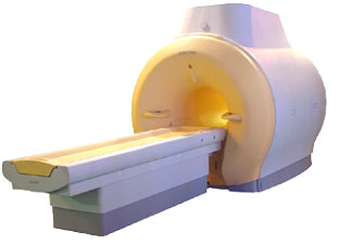
CHORUS 1.5T™InfoSheet: - Devices -
Intro, 
Types of Magnets, 
Overview, 
etc.MRI Resource Directory:
 - Devices -
 
www.isoltech.co.kr/english/product/default.htm 'Next generation MRI system 1.5T CHORUS developed by ISOL Technology is optimized for both clinical diagnostic imaging and for research development.
CHORUS offers the complete range of feature oriented advanced imaging techniques- for both clinical routine and research. The compact short bore magnet, the patient friendly design and the gradient technology make the innovation to new degree of perfection in magnetic resonance.'
Device Information and Specification
CLINICAL APPLICATION
Whole body
CONFIGURATION
Short bore
Head, C-spine, L-spine, TMJ, Knee, Shoulder, General purpose, Phased Array System: 4 digital receiver channels (Up to 12 channels)
Optional
SYNCHRONIZATION
ECG/peripheral: Optional/yes, respiratory gating
PULSE SEQUENCES
Spin Echo, Gradient Echo, Fast Spin Echo, Inversion Recovery (STIR, Fluid Attenuated Inversion Recovery), FLASH, FISP, PSIF, Turbo Flash ( MPRAGE ),TOF MR Angiography, Standard echo planar imaging package (SE-EPI, GE-EPI), Optional: Advanced P.A. Imaging Package (up to 4 ch.), Advanced echo planar imaging package, Single Shot and Diffusion Weighted EPI, IR/FLAIR EPI
IMAGING MODES
2D/3D, Travelling Sat, Multi-Slab 3D, MTC and TONE Pulse Sequence, Fat/Water Suppression, Presaturation (up to 6 bands), Flow Compensation using GMR pulse, Multi-Slice, Multi-Group Imaging
SINGLE/MULTI SLICE
Image reconstruction time (2562 ) : 0.02 s
FOV
40 cm
BORE DIAMETER
or W x H
58 cm diameter (patient)
MAGNET WEIGHT
4050 kg
H*W*D
233 x 206 x 160 cm
COOLING SYSTEM TYPE
Water-cooled coil and air-cooled amplifier
CRYOGEN USE
0.07 L/hr helium
STRENGTH
20 mT/m (Upto 27 mT/m)
5-GAUSS FRINGE FIELD
2.5 m / 3.8 m
Passive and active
spacer

• View the DATABASE results for 'CHORUS 1.5T™' (2).Open this link in a new window

Searchterm 'Gradient Magnetic Field' was also found in the following services: 
spacer
News  (1)  Resources  (3)  
 
ENCORE 0.5T™InfoSheet: - Devices -
Intro, 
Types of Magnets, 
Overview, 
etc.MRI Resource Directory:
 - Devices -
 
www.isoltech.co.kr/english/product/05t.htm 'MRI system is not an expensive equipment anymore. ENCORE developed by ISOL Technology is a low cost MRI system with the advantages like of the 1.0T MRI scanner. Developed specially for the overseas market, the ENCORE is gaining popularity in the domestic market by medium sized hospitals.
Due to the optimum RF and Gradient application technology. ENCORE enables to obtain high resolution imaging and 2D/3D Angio images which was only possible in high field MR systems.'
- Less consumption of the helium gas due to the ultra-lightweight magnet specially designed and manufactured for ISOL. - Cost efficiency MR system due to air cooling type (equivalent to permanent magnetic). - Patient processing speed of less than 20 minutes.'
Device Information and Specification
CLINICAL APPLICATION
Whole body
CONFIGURATION
Short bore compact
Head, C-spine, L-spine, TMJ, Knee, Shoulder, General purpose, Phased Array System: 4 digital receiver channels (Up to 12 channels)
SYNCHRONIZATION
ECG/peripheral: Optional/yes, respiratory gating
SINGLE/MULTI SLICE
Image reconstruction time (2562 ) : 0.02 s
FOV
40 cm
BORE DIAMETER
or W x H
58 cm diameter
MAGNET WEIGHT
3200 kg
H*W*D
200 x 168 x 187 cm
COOLING SYSTEM TYPE
Air-cooled coil and amplifier
CRYOGEN USE
0.05 L/hr helium
STRENGTH
15 mT/m
5-GAUSS FRINGE FIELD
2.3 m / 3.1 m
Passive and active
spacer

• View the DATABASE results for 'ENCORE 0.5T™' (2).Open this link in a new window

MRI Resources 
Diffusion Weighted Imaging - RIS - Developers - DICOM - Hospitals - Cardiovascular Imaging
 
Lung ImagingMRI Resource Directory:
 - Lung Imaging -
 
Lung imaging is furthermore a challenge in MRI because of the predominance of air within the lungs and associated susceptibility issues as well as low signal to noise of the inflated lung parenchyma. Cardiac and respiratory triggered or breath hold sequences allow diagnostic imaging, however a comparable image quality with computed tomography is still difficult to achieve.
Assumptions for lung MRI:
•
Low signal to noise ratio of the inherently low lung proton density.
•
Cardiac and respiratory motion artifacts.
•
Magnetic susceptibility effects of large magnetic field gradients.
•
Very short transverse relaxation times and significant diffusion yielding short T2 (30-70 msec), short T2* (1-3 msec), and additional long T1 relaxation times (1300-1500 msec).
•
The extreme short T2 values are responsible for a fast signal decay during a single shot readout, resulting in blurring.

The current trends in MRI are the use of new imaging technologies and increasingly powerful magnetic fields. Among these technologies are parallel imaging techniques as well as ventilation agents like hyperpolarized helium for the use as an inert inhalational contrast agent to study lung ventilation properties. With hyperpolarized gases clear images of the lungs can be obtained without using a large magnetic field (see also back projection imaging). Single shot sequences (e.g. TSE or Half Fourier Acquisition Single Shot Turbo Spin Echo HASTE) used in lung MR imaging benefits from parallel imaging techniques due to reduced relaxation time effects during the echo train and therefore reduced image blurring as well as reduced motion artifacts.
In the future, more effective contrast agents may provide an alternative solution to the need for high field MRI. Dynamic contrast enhanced MRI perfusion has demonstrated a potential in the diagnosis of pulmonary embolism or to characterize lung cancer and mediastinal tumors. 3D contrast enhanced magnetic resonance angiography of the thoracic vessel.

See also the related poll result: 'MRI will have replaced 50% of x-ray exams by'
 
Images, Movies, Sliders:
 Anatomic Imaging of the Lungs  Open this link in a new window
      

Courtesy of  Robert R. Edelman
 Normal Lung Gd Perfusion MRI  Open this link in a new window
      

Courtesy of  Robert R. Edelman

 MRI Thorax Basal Plane  Open this link in a new window
 
Radiology-tip.comradLung Scintigraphy
spacer

• View the DATABASE results for 'Lung Imaging' (7).Open this link in a new window


• View the NEWS results for 'Lung Imaging' (3).Open this link in a new window.
 
Further Reading:
  Basics:
A safer approach for diagnostic medical imaging
Monday, 29 September 2014   by www.eurekalert.org    
Parallel Lung Imaging(.pdf)
  News & More:
Chest MRI a viable alternative to chest CT in COVID-19 pneumonia follow-up
Monday, 21 September 2020   by www.healthimaging.com    
CT Imaging Features of 2019 Novel Corona virus (2019-nCoV)
Tuesday, 4 February 2020   by pubs.rsna.org    
Polarean Imaging Phase III Trial Results Point to Potential Improvements in Lung Imaging
Wednesday, 29 January 2020   by www.diagnosticimaging.com    
Low Power MRI Helps Image Lungs, Brings Costs Down
Thursday, 10 October 2019   by www.medgadget.com    
Chest MRI Using Multivane-XD, a Novel T2-Weighted Free Breathing MR Sequence
Thursday, 11 July 2019   by www.sciencedirect.co    
Researchers Review Importance of Non-Invasive Imaging in Diagnosis and Management of PAH
Wednesday, 11 March 2015   by lungdiseasenews.com    
New MRI Approach Reveals Bronchiectasis' Key Features Within the Lung
Thursday, 13 November 2014   by lungdiseasenews.com    
MRI techniques improve pulmonary embolism detection
Monday, 19 March 2012   by medicalxpress.com    
  News & More:
Partnership with VIDA to streamline adoption of advanced MRI of the lungs
Monday, 11 September 2023   by www.itnonline.com    
MRI Resources 
Pacemaker - Supplies - Directories - Lung Imaging - Implant and Prosthesis - Liver Imaging
 
previous      16 - 20 (of 27)     next
Result Pages : [1]  [2]  [3 4 5 6]
 Random Page
 
Share This Page
FacebookTwitterLinkedIn

MR-TIP    
Community   
User
Pass
Forgot your UserID/Password ?    


How AI will impact MRI :
only diagnostics 
saving time 
reducing cost 
makes planning obsolete 
reduce human knowledge 
not at all 

Look
      Ups


MR-TIP.com uses cookies! By browsing MR-TIP.com, you agree to our use of cookies.

Magnetic Resonance - Technology Information Portal
Member of SoftWays' Medical Imaging Group - MR-TIP • Radiology-TIP • Medical-Ultrasound-Imaging • 
Copyright © 2003 - 2024 SoftWays. All rights reserved. [ 29 April 2024]
Terms of Use | Privacy Policy | Advertising
 [last update: 2024-02-26 03:41:00]