 | Info
Sheets |
| | | | | | | | | | | | | | | | | | | | | | | | |
 | Out-
side |
| | | | |
|
| | | | |
Result : Searchterm 'Dixon' found in 1 term [ ] and 11 definitions [ ] and 11 definitions [ ] ]
| | previous 6 - 10 (of 12) nextResult Pages : [1] [2 3] |  | |  | Searchterm 'Dixon' was also found in the following services: | | | | |
| |  |
| |
|
Liver imaging can be performed with sonography, computed tomography (CT) and magnetic resonance imaging ( MRI). Ultrasound is, caused by the easy access, still the first-line imaging method of choice; CT and MRI are applied whenever ultrasound imaging yields vague results. Indications are the characterization of metastases and primary liver tumors e.g., benign lesions such as focal nodular hyperplasia (FNH), adenoma, hemangioma and malignant lesions (cancer) such as hepatocellular carcinomas (HCC).
The decision, which medical imaging modality is more suitable, MRI or CT, is dependent on the different factors. CT is less costly and more widely available; modern multislice scanners provide high spatial resolution and short scan times but has the disadvantage of radiation exposure.
With the introduction of high performance MR systems and advanced sequences the image quality of MRI for the liver has gained substantially. Fast spin echo or single shot techniques, often combined with fat suppression, are the most common T2 weighted sequences used in liver MRI procedures.
Spoiled gradient echo sequences are used as ideal T1 weighted sequences for evaluating of the liver. The repetition time (TR) can be sufficiently long to acquire enough sections covering the entire liver in one pass, and to provide good signal to noise. The TE should be the shortest in phase echo time (TE), which provides strong T1 weighting, minimizes magnetic susceptibility effects, and permits acquisition within one breath hold to cover the whole liver. A flip angle of 80° provides good T1 weighting and less of power deposition and tissue saturation than a larger flip angle that would provide comparable T1 weighting.
Liver MRI is very dependent on the administration of contrast agents, especially when detection and characterization of focal lesions are the issues. Liver MRI combined with MRCP is useful to evaluate patients with hepatic and biliary disease.
Gadolinium chelates are typical non-specific extracellular agents diffusing rapidly to the extravascular space of tissues being cleared by glomerular filtration at the kidney. These characteristics are somewhat problematic when a large organ with a huge interstitial space like the liver is imaged. These agents provide a small temporal imaging window (seconds), after which they begin to diffuse to the interstitial space not only of healthy liver cells but also of lesions, reducing the contrast gradient necessary for easy lesion detection. Dynamic MRI with multiple phases after i.v. contrast media (Gd chelates), with arterial, portal and late phase images (similar to CT) provides additional information.
An additional advantage of MRI is the availability of liver-specific contrast agents (see also Hepatobiliary Contrast Agents). Gd-EOB-DTPA (gadoxetate disodium, Gadolinium ethoxybenzyl dimeglumine, EOVIST Injection, brand name in other countries is Primovist) is a gadolinium-based MRI contrast agent approved by the FDA for the detection and characterization of known or suspected focal liver lesions.
Gd-EOB-DTPA provides dynamic phases after intravenous injection, similarly to non-specific gadolinium chelates, and distributes into the hepatocytes and bile ducts during the hepatobiliary phase. It has up to 50% hepatobiliary excretion in the normal liver.
Since ferumoxides are not eliminated by the kidney, they possess long plasmatic half-lives, allowing circulation for several minutes in the vascular space. The uptake process is dependent on the total size of the particle being quicker for larger particles with a size of the range of 150 nm (called superparamagnetic iron oxide). The smaller ones, possessing a total particle size in the order of 30 nm, are called ultrasmall superparamagnetic iron oxide particles and they suffer a slower uptake by RES cells. Intracellular contrast agents used in liver MRI are primarily targeted to the normal liver parenchyma and not to pathological cells. Currently, iron oxide based MRI contrast agents are not marketed.
Beyond contrast enhanced MRI, the detection of fatty liver disease and iron overload has clinical significance due to the potential for evolution into cirrhosis and hepatocellular carcinoma. Imaging-based liver fat quantification (see also Dixon) provides noninvasively information about fat metabolism; chemical shift imaging or T2*-weighted imaging allow the quantification of hepatic iron concentration.
See also Abdominal Imaging, Primovistâ„¢, Liver Acquisition with Volume Acquisition (LAVA), T1W High Resolution Isotropic Volume Examination (THRIVE) and Bolus Injection.
For Ultrasound Imaging (USI) see Liver Sonography at Medical-Ultrasound-Imaging.com. | | | | | | | | | | | | | | | | | |  Further Reading: Further Reading: | | Basics:
|
|
News & More:
|  |
Utility and impact of magnetic resonance elastography in the clinical course and management of chronic liver disease
Saturday, 20 January 2024 by www.nature.com | | |
Even early forms of liver disease affect heart health, Cedars-Sinai study finds
Thursday, 8 December 2022 by www.eurekalert.org | | |
For monitoring purposes, AI-aided MRI does what liver biopsy does with less risk, lower cost
Wednesday, 28 September 2022 by radiologybusiness.com | | |
Perspectum: High Liver Fat (Hepatic Steatosis) Linked to Increased Risk of Hospitalization in COVID-19 Patients With Obesity
Monday, 29 March 2021 by www.businesswire.com | | |
EMA's final opinion confirms restrictions on use of linear gadolinium agents in body scans
Friday, 21 July 2017 by www.ema.europa.eu | | |
T2-Weighted Liver MRI Using the MultiVane Technique at 3T: Comparison with Conventional T2-Weighted MRI
Friday, 16 October 2015 by www.ncbi.nlm.nih.gov | | |
EORTC study aims to qualify ADC as predictive imaging biomarker in preoperative regimens
Monday, 4 January 2016 by www.eurekalert.org | | |
MRI effectively measures hemochromatosis iron burden
Saturday, 3 October 2015 by medicalxpress.com | | |
Total body iron balance: Liver MRI better than biopsy
Sunday, 15 March 2015 by www.eurekalert.org |
|
| |
| | | | | |
| |
|
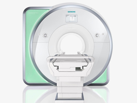
From Siemens Medical Systems;
Received FDA clearance in 2010.
The MAGNETOM Aera is a patient friendly, comfortable 1.5 Tesla MRI system with advanced radio frequency chain.
The system is equipped with the Tim 4G and Dot system (Total imaging matrix + Day optimizing throughput), to enhance both productivity and image quality.
Tim 4G technology provides improved SNR. The standard system configuration of 48 radio frequency channels and 204 coil elements creates an imaging matrix that allows maximum use of coil elements at full field of view. Dot provides improved image consistency through new features like auto align, auto FoV and automatic bolus detection.
Device Information and Specification
CLINICAL APPLICATION
Whole body
Head, spine, torso/ body coil, neurovascular, cardiac, neck, shoulder, knee, wrist, foot//ankle and multi-purpose flex coils. Peripheral vascular, breast, shoulder. Up to 60% more SNR with Tim 4G.
CHANNELS (min. / max. configuration)
48, 64
MINIMUM TE
3-D GRE: 0.22 (256 matrix), Ultra-short TE
At isocenter: L-R 70 cm, A-P (with table) 55 cm
MAGNET WEIGHT (gantry included)
3121 kg
DIMENSION H*W*D (gantry included)
145 x 231 x 219 cm
MAX. AMPLITUDE
33 or 45 mT/m
3 linear with 20 coils, 5 nonlinear 2nd-order
POWER REQUIREMENTS
380 / 400 / 420 / 440 / 460 / 480 V, 3-phase + ground; 85 kVA
| | | | | |
| | | | | |
| |
|
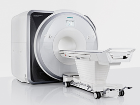
From Siemens Medical Systems;
Received FDA clearance in 2013.
The MAGNETOM Prisma is the 3T PowerPack for exploration that offers most demanding clinical and research challenges of today and the future. The latest parallel transmit technology, TimTX TrueShape, enables zooming into specific body regions for enhanced image quality. Furthermore, the Tim 4G integrated coil technology offers remarkable imaging flexibility and supports complex
examinations across the whole body.
Onsite upgrades to the MAGNETOM Prisma for customers who have already installed the 3 Tesla MAGNETOM Trio are possible.
Device Information and Specification
CLINICAL APPLICATION
Whole Body
CONFIGURATION
Ultra-short bore
Head, spine, torso/ body coil, neurovascular, cardiac, neck, shoulder, knee, wrist, foot//ankle and multi-purpose flex coils. Peripheral vascular, breast, shoulder.
CHANNELS (min. / max. configuration)
64, 128
MAGNET WEIGHT (gantry included)
13000 kg
DIMENSION H*W*D (gantry included)
173 x 230 x 222 cm
Passive, active; first order,
second order
POWER REQUIREMENTS
380 / 400 / 420 / 440 / 460 / 480 V, 3-phase + ground;
| | | | | |
| | | Searchterm 'Dixon' was also found in the following services: | | | | |
| | |
| |
|
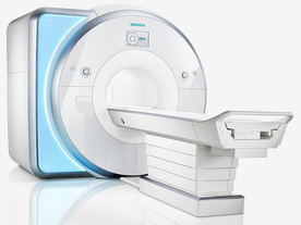
From Siemens Medical Systems;
Received FDA clearance in 2010.
MAGNETOM Skyra is a top-of-the-line, patient friendly wide bore 3 Tesla MRI system.
The system is equipped with the Tim 4G and Dot system (Total imaging matrix and Day optimizing throughput), to enhance both productivity and image quality with the complete range of advanced applications for clinical routine and research. Tim 4G features lighter, trimmer MRI coils that take up less space inside the magnet but deliver a high coil element density with increased signal to noise ratio and the possibility to use high iPAT factors.
Device Information and Specification
CLINICAL APPLICATION
Whole Body
Head, spine, torso/ body coil, neurovascular, cardiac, neck, shoulder, knee, wrist, foot//ankle and multi-purpose flex coils. Peripheral vascular, breast, shoulder.
CHANNELS (min. / max. configuration)
48, 64, 128
Chemical shift imaging, single voxel spectroscopy
MINIMUM TE
3D T1 spoiled GRE: 0.22 (256 matrix), Ultra-short TE
At isocenter: L-R 70 cm, A-P (with table) 55 cm
MAGNET WEIGHT (gantry included)
5768 kg
DIMENSION H*W*D (gantry included)
173 x 231 x 219 cm
COOLING SYSTEM
Water; single cryogen, 2 stage refrigeration
3 linear with 20 coils, 5 nonlinear 2nd-order
POWER REQUIREMENTS
380 / 400 / 420 / 440 / 460 / 480 V, 3-phase + ground; 110 kVA
| | | | | |
| | | | | |
| |
|
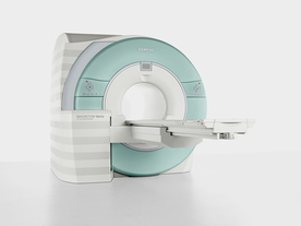
From Siemens Medical Systems;
Received FDA clearance in 2007.
The MAGNETOM Verio provides up to 102 integrated matrix coil elements and up to 32 independent radiofrequency channels that allow flexible coil combinations to make patient and coil repositioning virtually unnecessary. The Tim (total imaging matrix) technology also increases patient throughput due to a shorter scan time.
The open bore design offers great comfort for patients of all shapes and sizes.
Device Information and Specification
CLINICAL APPLICATION
Whole Body
CONFIGURATION
Ultra-short open bore
Head, spine, torso/ body coil, neurovascular, cardiac, neck and multi-purpose flex coils. Peripheral vascular, breast, shoulder, knee, wrist, foot//ankle, TMJ optional.
CHANNELS (min. / max. configuration)
8, 18, 32
Chemical shift imaging, single voxel spectroscopy
MAGNET WEIGHT (gantry included)
8200 kg
DIMENSION H*W*D (gantry included)
173 x 230 x 222 cm
Passive, active; first order,
second order standard
POWER REQUIREMENTS
380 / 400 / 420 / 440 / 460 / 480 V, 3-phase + ground; 110 kVA
| | | | | |
| | | | |
| | | |
|
| |
 | Look
Ups |
| |


