 | Info
Sheets |
| | | | | | | | | | | | | | | | | | | | | | | | |
 | Out-
side |
| | | | |
|
| | | | |
Result : Searchterm 'Dedicated extremity' found in 0 term [ ] and 9 definitions [ ] and 9 definitions [ ], (+ 2 Boolean[ ], (+ 2 Boolean[ ] results ] results
| | previous 6 - 10 (of 11) nextResult Pages : [1 2] [3] |  | |  | Searchterm 'Dedicated extremity' was also found in the following services: | | | | |
| |  |
| |
|
| | | | | | | | | | |  Further Reading: Further Reading: | | Basics:
|
|
News & More:
|  |
Safety of Bedside Portable Low-Field Brain MRI in ECMO Patients Supported on Intra-Aortic Balloon Pump
Friday, 18 November 2022 by www.mdpi.com | | |
Researchers at the University of Tsukuba develop a portable MRI system specifically for identifying wrist cartilage damage among athletes, providing a convenient means of early detection and treatment of injuries
Tuesday, 26 April 2022 by www.tsukuba.ac.jp | | |
This bizarre looking helmet can create better brain scans
Friday, 11 February 2022 by www.sciencedaily.com | | |
A low-cost and shielding-free ultra-low-field brain MRI scanner
Tuesday, 14 December 2021 by www.nature.com | | |
Portable MRI provides life-saving information to doctors treating strokes
Thursday, 5 August 2021 by news.yale.edu | | |
Synaptive Evry, an MRI for Any Space, Cleared by FDA
Thursday, 30 April 2020 by www.medgadget.com | | |
World's First Portable MRI Cleared by FDA
Monday, 17 February 2020 by www.medgadget.com | | |
Introducing a point-of-care MRI system
Tuesday, 29 October 2019 by healthcare-in-europe.com | | |
Opportunities in Interventional and Diagnostic Imaging by Using High-performance Low-Field-Strength MRI
Tuesday, 1 October 2019 by pubs.rsna.org | | |
Portable 'battlefield MRI' comes out of the lab
Thursday, 30 April 2015 by physicsworld.com | | |
Portable MRI could aid wounded soldiers and children in the third world
Thursday, 23 April 2015 by phys.org |
|
| |
| | | | | |
| |
|
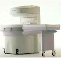
From Siemens Medical Systems;
This open MRI system is a dedicated extremity scanner, which is easy to use and easy to install. Whether you want to use it as a supplement to your whole-body scanner or you want to specialize in orthopedic exams,
MAGNETOM Jazz™ offers you a cost effective opportunity to make the most of your capabilities.
Device Information and Specification GRE, IR, FIR, STIR, TrueIR/FISP, FSE, MT, SS-FSE, MT-SE, MTC, MSE, GMR, fat/water sat./exc. IMAGING MODES Single, multislice, volume study, multi angle 512 x 512 full screen display POWER REQUIREMENTS 380/400/420/440/480 V | | | |
• View the DATABASE results for 'MAGNETOM Jazz™' (2).
| | | | |
| | | | | |
| |
|

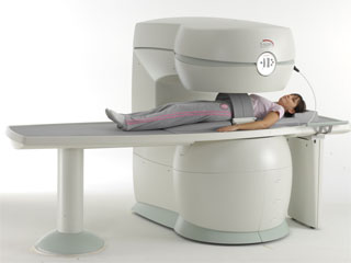
From ONI Medical Systems, Inc.;
MSK-Extreme™ MRI system is a dedicated high field extremity imaging device, designed to provide orthopedic surgeons and other physicians with detailed diagnostic images of the foot, ankle, knee, hand, wrist and elbow, all with the clinical confidence and advantages derived from high field, whole body MRI units. The light weight (less than 650 kg) of the OrthOne System performs rapid patient studies, is easy to operate, has a patient friendly open environment and can be installed in a practice office or hospital, all at a cost similar to a low field extremity machine.
New features include a more powerful operating system that offers increased scan speed as well as a 160-mm knee coil with higher signal to noise ratio, and the option of a CD burner.
Device Information and Specification 16 cm knee, 18 cm lower extremity;; 12.3 cm upper extremity, additional high resolution v-SPEC Coils: 80 mm, 100 mm, or 145 mm. SE, FSE, GE2D, GE3D, Inversion recovery (IR), Driven Equilibrium, Fat Saturation (FS), STIR, MT, PD, Flow Compensation (FC), RF spoiling, MTE, No Phase Wrap (NPW) IMAGING MODES Scout, single, multislice, volume 2D less than 200 msec/image X/Y: 64-512; 2 pixel steps 4,096 grey lvls; 256 lvls in 3D POWER REQUIREMENTS 115VAC, 1phase, 20A; 208VAC, 3 phase, 30A COOLING SYSTEM TYPE LHe with 2 stage cold head 1.25m radial x 1.8m axial | | | | | | | Further Reading: | Basics:
|
|
| |
| | | Searchterm 'Dedicated extremity' was also found in the following services: | | | | |
| | |
| |
|
From MagneVu;
The MagneVu 1000 is a compact, robust, and portable, permanent magnet MRI system and operates without special shielding or costly site preparation.
This MRI device utilizes a patented non-homogeneous magnetic field image acquisition method to achieve high performance imaging. The MagneVu 1000 MRI scanner is designed for MRI of the extremities with the current specialty areas in diabetes and rheumatoid arthritis. Easy access is afforded for claustrophobic, pediatric, or limited mobility patients. In August 1998
FDA marketing clearance and other regulatory approvals have been received. Until 2008, over 130 devices in the US are in use. Some further developments of MagneVu's extremity scanner are: 'truly Plug n' Play MRI™' and iSiS ( which adds wireless capability to the second generation MV1000-XL).
Device Information and Specification IMAGING MODES 3-dimensional multi-echo data acquisition | | | |
• View the DATABASE results for 'MagneVu 1000' (3).
| | | | | | Further Reading: | News & More:
|
|
| |
| | | | |  |
| |
|
From Esaote S.p.A.;
Esaote introduced the S-SCAN at RSNA in November 2007. The S-SCAN is a dedicated joint and spine MR scanner derived from the company's earlier G-SCAN system. Unlike the G-SCAN, neither the patient table nor
the magnet can rotate from horizontal to vertical position. The patient table can only moved manually. Improved electronics, new coils for lumbar and cervical spine, new pulse sequences, a modified version of the magnet poles and gradient coils are used with a new software release in the S-SCAN.
Esaote North America is the exclusive U.S. distributor of this MRI device.
Device Information and Specification SE, GE, IR, STIR, TSE, 3D CE, GE-STIR, 3D GE, ME, TME, HSE POWER REQUIREMENTS 3 kW; 110/220 V single phase | | | |
• View the DATABASE results for 'S-SCAN' (3).
| | |
• View the NEWS results for 'S-SCAN' (1).
| | | | | | Further Reading: | News & More:
|
|
| |
| | | | |
| | | |
|
| |
 | Look
Ups |
| |


