Magnetic Resonance - Technology Information Portal Welcome to MRI Technology
Info
  Sheets

Out-
      side


 'Slice' 
SEARCH FOR    
 
  2 3 5 A B C D E F G H I J K L M N O P Q R S T U V W X Y Z
Result : Searchterm 'Slice' found in 19 terms [] and 159 definitions []
previous     56 - 60 (of 178)     next
Result Pages : [1 2 3 4]  [5 6 7 8 9 10 11 12 13 14 15 16 17 18 19 20 ... ]
Searchterm 'Slice' was also found in the following services: 
spacer
News  (6)  Resources  (5)  Forum  (25)  
 
Cardiac Gating
 
This method synchronize the heartbeat with the beginning of the TR, whereat the r wave is used as the trigger. Cardiac gating times the acquisition of MR data to physiological motion in order to minimize motion artifacts. ECG gating techniques are useful whenever data acquisition is too slow to occur during a short fraction of the cardiac cycle.
Image blurring due to cardiac-induced motion occurs for imaging times of above approximately 50 ms in systole, while for imaging during diastole the critical time is of the order of 200-300 ms. The acquisition of an entire image in this time is only possible with using ultrafast MR imaging techniques. If a series of images using cardiac gating or real-time echo planar imaging EPI are acquired over the entire cardiac cycle, pixel-wise velocity and vascular flow can be obtained.
In simple cardiac gating, a single image line is acquired in each cardiac cycle. Lines for multiple images can then be acquired successively in consecutive gate intervals. By using the standard multiple slice imaging and a spin echo pulse sequence, a number of slices at different anatomical levels is obtained. The repetition time (TR) during a ECG-gated acquisition equals the RR interval, and the RR interval defines the minimum possible repetition time (TR). If longer TRs are required, multiple integers of the RR interval can be selected. When using a gradient echo pulse sequence, multiple phases of a single anatomical level or multiple slices at different anatomical levels can be acquired over the cardiac cycle.
Also called cardiac triggering.
 
Images, Movies, Sliders:
 Cardiac Infarct Short Axis Cine Overview  Open this link in a new window
    

Courtesy of  Robert R. Edelman
 Infarct 4 Chamber Cine  Open this link in a new window
    

Courtesy of  Robert R. Edelman
 
spacer
 
• Related Searches:
    • Heart MRI
    • Cardiac MRI
    • Cerebro Spinal Fluid Pulsation Artifact
    • Cardiovascular Imaging
    • Arrhythmia Rejection
 
Further Reading:
  Basics:
Cardiac MRI - Technical Aspects Primer
Wednesday, 7 August 2002
Electrocardiogram in an MRI Environment: Clinical Needs, Practical Considerations, Safety Implications, Technical Solutions and Future Directions
Wednesday, 25 January 2012   by cdn.intechopen.com    
Motion-compensation of Cardiac Perfusion MRI using a Statistical Texture Ensemble(.pdf)
June 2003   by www.imm.dtu.dk    
Searchterm 'Slice' was also found in the following services: 
spacer
Radiology  (24) Open this link in a new windowUltrasound  (9) Open this link in a new window
Excelart AG™ with PianissimoInfoSheet: - Devices -
Intro, 
Types of Magnets, 
Overview, 
etc.MRI Resource Directory:
 - Devices -
 
http://www.medical.toshiba.com/clinical/radiology/15texcelart.htm From Toshiba America Medical Systems Inc.;
the EXCELART is a superconducting whole body MRI system with a short wide-bore magnet, operating at 1.5 T. It features powerful high-speed gradients with a revolutionary gradient acoustic noise reduction system: Pianissimo. The dramatic reduction of gradient acoustic noise by Pianissimo greatly enhances patient comfort during exams. The standard array platform and a wide range of array coils ensure excellent images. A powerful 64-bit RISC-based computer system and newly developed array processor realize high productivity.
Device Information and Specification
CLINICAL APPLICATION
Whole body
CONFIGURATION
Cylindrical Wide Short Bore
Opt. (WIP) Single and Multi Voxel
SYNCHRONIZATION
ECG: standard; peripheral: optional, respiratory gating
PULSE SEQUENCES
SE, FE, IR, FastSE, FastIR, FastFLAIR, Fast STIR, FastFE, FASE, Hybrid EPI, Multi Shot EPI; Angiography: 2D(gate/non-gate)/3D TOF, SORS-STC
IMAGING MODES
Single, multislice, volume study
TR
3.5-30000 msec
TE
8 msec min. SE; 1.2 msec min. FE
SINGLE/MULTI SLICE
less than 0.015 (256x256)
FOV
2 cm
1.0 min. 2-DFT: 0.2 min. 3-DFT
Up to 1024
MEASURING MATRIX
32-1024, phase;; 64-1024, freq.
PIXEL INTENSITY
256 gray levels
BORE DIAMETER
or W x H
65.5 cm, patient aperture
MAGNET WEIGHT
4050 kg (bare magnet incl. L-He)
H*W*D
235 x 219 x 199 cm
COOLING SYSTEM TYPE
Closed-loop water-cooled
CRYOGEN USE
Liquid helium: approx. less than 0.05 L/hr
STRENGTH
25 mT/m
5-GAUSS FRINGE FIELD
2.5 m / 4.0 m
Passive, active, auto-active
spacer

• View the DATABASE results for 'Excelart AG™ with Pianissimo' (2).Open this link in a new window

MRI Resources 
Colonography - Image Quality - Libraries - Musculoskeletal and Joint MRI - MR Myelography - Service and Support
 
Excelart XG™ with PianissimoInfoSheet: - Devices -
Intro, 
Types of Magnets, 
Overview, 
etc.MRI Resource Directory:
 - Devices -
 
http://www.medical.toshiba.com/clinical/radiology/15texcelart.htm From Toshiba America Medical Systems Inc.;
the EXCELART is a superconducting whole body MRI system with a short wide-bore magnet, operating at 1.5 T. It features powerful high-speed gradients with a revolutionary gradient acoustic noise reduction system: Pianissimo. The dramatic reduction of gradient acoustic noise by Pianissimo greatly enhances patient comfort during exams. The standard array platform and a wide range of array coils ensure excellent images. A powerful 64-bit RISC-based computer system and newly developed array processor realize high productivity.
Device Information and Specification
CLINICAL APPLICATION
Whole body
CONFIGURATION
Cylindrical Wide Short Bore
Optional (WIP)
SYNCHRONIZATION
ECG/peripheral, respiratory gating
PULSE SEQUENCES
SE, FE, IR, FastSE, FastIR, FastFLAIR, Fast STIR, FastFE, FASE, Hybrid EPI, Multi Shot EPI; Angiography: 2D(gate/non-gate)/3D TOF, SORS-STC
IMAGING MODES
Single, multislice, volume study
TR
2.6-30000 msec
TE
8 msec min. SE; 0.9 msec min. FE
SINGLE/MULTI SLICE
less than 0.011 (256x256)
FOV
2 cm
1.0 min. 2-DFT: 0.2 min. 3-DFT
Up to 1024
MEASURING MATRIX
32-1024, phase;; 64-1024, freq.
PIXEL INTENSITY
256 gray levels
BORE DIAMETER
or W x H
65.5 cm, patient aperture
MAGNET WEIGHT
4050 kg (bare magnet incl. L-He)
H*W*D
235 x 219 x 199 cm
POWER REQUIREMENTS
380/400/415/440/480 V
COOLING SYSTEM TYPE
Closed-loop water-cooled
CRYOGEN USE
Liquid helium: approx. less than 0.05 L/hr
STRENGTH
25 mT/m
5-GAUSS FRINGE FIELD
2.5 m / 4.0 m
Passive, active, auto-active
spacer

• View the DATABASE results for 'Excelart XG™ with Pianissimo' (2).Open this link in a new window

 
Further Reading:
  News & More:
Angio, cardiac imaging top list for MR and CT
1999   by www.diagnosticimaging.com    
Searchterm 'Slice' was also found in the following services: 
spacer
News  (6)  Resources  (5)  Forum  (25)  
 
MAGNETOM Allegra™InfoSheet: - Devices -
Intro, 
Types of Magnets, 
Overview, 
etc.MRI Resource Directory:
 - Devices -
 
www.med.siemens.com/med/d/gg/mr/products/allegra.htm From Siemens Medical Systems;
the 3 T MAGNETOM Allegra is a dedicated MR headscanner, perfect as a research system in cognitive and neuroscience with MRS and fMRI. MAGNETOM Allegra is a full member of the MAGNETOM product family. It uses many common components, i.e. electronics, computer system, software and pulse sequence concepts.
Device Information and Specification
CLINICAL APPLICATION
CONFIGURATION
Short bore
Yes/Mutli-nuclear MRS
PULSE SEQUENCES
GRE, IR, FIR, STIR, TrueIR/FISP, FSE, FLAIR, MT, SS-FSE, MT-SE, MTC, MSE, EPI, GMR, fat/water sat./exc.
IMAGING MODES
Single, multislice, volume study, multi angle, multi oblique
SINGLE/MULTI SLICE
178 images/sec at 256 x 256 at 100% FOV
FOV
22 cm
Min 2D/3D: 0.1/0.05 mm
1024 x 1024 full screen display
MEASURING MATRIX
64 x 64 to 1024 x 1024
10 micrometer
BORE DIAMETER
or W x H
60 x 60 cm
MAGNET WEIGHT (gantry included)
5500 kg
DIMENSION H*W*D (gantry included)
220 x 220 x 147 cm
POWER REQUIREMENTS
380/400/420/440/480 V
COOLING SYSTEM TYPE
Single cryogen, 2 stage refrig.
CRYOGEN USE
0.1 L/hr helium
STRENGTH
40 mT/m
Passive, act.; 1st order std./2nd opt.
spacer

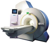
• View the DATABASE results for 'MAGNETOM Allegra™' (2).Open this link in a new window

Searchterm 'Slice' was also found in the following services: 
spacer
Radiology  (24) Open this link in a new windowUltrasound  (9) Open this link in a new window
MAGNETOM Harmony™InfoSheet: - Devices -
Intro, 
Types of Magnets, 
Overview, 
etc.MRI Resource Directory:
 - Devices -
 
www.med.siemens.com/med/d/gg/mr/products/harmony.htm From Siemens Medical Systems;
the MAGNETOM Harmony system is a high-performance MR imaging system based on 1.0 T field strength for best clinical routine MRI. It combines patient-friendly design features with the benefits of short imaging time, high anatomical resolution, and highest productivity.
Device Information and Specification
CLINICAL APPLICATION
Whole body
CONFIGURATION
Compact
Body, head, spine, knee, neck, TMJ, extremity, breast, shoulder, others
Yes/SVS opt.
SYNCHRONIZATION
ECG/peripheral, respiratory gating
PULSE SEQUENCES
GRE, IR, FIR, STIR, TrueIR/FISP, FSE, FLAIR, MT, SS-FSE, MT-SE, MTC, MSE, EPI, GMR, fat/water sat./exc.
IMAGING MODES
Single, multislice, volume study, multi angle, multi oblique
TR
2.4 msec std.; 2.0 opt.; 1.8 w/ 30mT/m at 256matrix
TE
1.1 msec std.; 0.9 opt.; 0.78 w/30 mT/m at 256matrix
SINGLE/MULTI SLICE
178 images/sec at 256 x 256 at 100% FOV
FOV
0.5 cm - 50 cm
Min 2D/3D: 0.1/0.05 mm
1024 x 1024 full screen display
MEASURING MATRIX
64 x 64 to 1024 x 1024
21 micrometer in plane, 11 micrometer optional
BORE DIAMETER
or W x H
60 cm
MAGNET WEIGHT (gantry included)
3500kg, 5000kg in operation
DIMENSION H*W*D (gantry included)
POWER REQUIREMENTS
380/400/420/440/480 V
COOLING SYSTEM TYPE
Single cryogen, 2 stage refrig.
CRYOGEN USE
Liquid He
STRENGTH
20/35 mT/m standard, 30/52 opt.
5-GAUSS FRINGE FIELD
2.5 m / 3.9 m
Passive, act.; 1st order std./2nd opt.
spacer

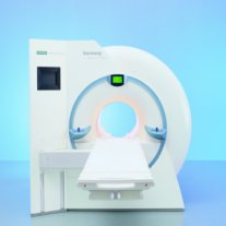
• View the DATABASE results for 'MAGNETOM Harmony™' (2).Open this link in a new window

 
Further Reading:
  Basics:
Harmony Brochure(.pdf)
MRI Resources 
Contrast Enhanced MRI - Intraoperative MRI - MRI Technician and Technologist Jobs - Hospitals - Contrast Agents - Shoulder MRI
 
previous      56 - 60 (of 178)     next
Result Pages : [1 2 3 4]  [5 6 7 8 9 10 11 12 13 14 15 16 17 18 19 20 ... ]
 Random Page
 
Share This Page
FacebookTwitterLinkedIn

MR-TIP    
Community   
User
Pass
Forgot your UserID/Password ?    


MRI is trending to low field magnets :
reduced costs will lead to this change 
AI will close the gap to high field 
only in remote areas 
is only temporary 
never 

Look
      Ups


MR-TIP.com uses cookies! By browsing MR-TIP.com, you agree to our use of cookies.

Magnetic Resonance - Technology Information Portal
Member of SoftWays' Medical Imaging Group - MR-TIP • Radiology-TIP • Medical-Ultrasound-Imaging • 
Copyright © 2003 - 2024 SoftWays. All rights reserved. [ 26 April 2024]
Terms of Use | Privacy Policy | Advertising
 [last update: 2024-02-26 03:41:00]