Magnetic Resonance - Technology Information Portal Welcome to MRI Technology
Info
  Sheets

Out-
      side


 'Resolution' 
SEARCH FOR    
 
  2 3 5 A B C D E F G H I J K L M N O P Q R S T U V W X Y Z
Result : Searchterm 'Resolution' found in 8 terms [] and 144 definitions []
previous     81 - 85 (of 152)     next
Result Pages : [1 2]  [3 4 5 6 7 8 9 10 11 12 13 14 15 16 17 18 19 20 ... ]
Searchterm 'Resolution' was also found in the following services: 
spacer
News  (40)  Resources  (13)  Forum  (15)  
 
Fast Spin EchoForum -
related threadsInfoSheet: - Sequences - 
Intro, 
Overview, 
Types of, 
etc.
 
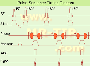
Fast Spin Echo Diagram (FSE) In the pulse sequence timing diagram, a fast spin echo sequence with an echo train length of 3 is illustrated. This sequence is characterized by a series of rapidly applied 180° rephasing pulses and multiple echoes, changing the phase encoding gradient for each echo.
The echo time TE may vary from echo to echo in the echo train. The echoes in the center of the K-space (in the case of linear k-space acquisition) mainly produce the type of image contrast, whereas the periphery of K-space determines the spatial resolution. For example, in the middle of K-space the late echoes of T2 weighted images are encoded. T1 or PD contrast is produced from the early echoes.
The benefit of this technique is that the scan duration with, e.g. a turbo spin echo turbo factor / echo train length of 9, is one ninth of the time. In T1 weighted and proton density weighted sequences, there is a limit to how large the ETL can be (e.g. a usual ETL for T1 weighted images is between 3 and 7). The use of large echo train lengths with short TE results in blurring and loss of contrast. For this reason, T2 weighted imaging profits most from this technique.
In T2 weighted FSE images, both water and fat are hyperintense. This is because the succession of 180° RF pulses reduces the spin spin interactions in fat and increases its T2 decay time. Fast spin echo (FSE) sequences have replaced conventional T2 weighted spin echo sequences for most clinical applications. Fast spin echo allows reduced acquisition times and enables T2 weighted breath hold imaging, e.g. for applications in the upper abdomen.
In case of the acquisition of 2 echoes this type of a sequence is named double fast spin echo / dual echo sequence, the first echo is usually density and the second echo is T2 weighted image. Fast spin echo images are more T2 weighted, which makes it difficult to obtain true proton density weighted images. For dual echo imaging with density weighting, the TR should be kept between 2000 - 2400 msec with a short ETL (e.g., 4).
Other terms for this technique are:
Turbo Spin Echo
Rapid Imaging Spin Echo,
Rapid Spin Echo,
Rapid Acquisition Spin Echo,
Rapid Acquisition with Refocused Echoes
 
Images, Movies, Sliders:
 Lumbar Spine T2 FSE Sagittal  Open this link in a new window
    

Courtesy of  Robert R. Edelman
 MRI - Anatomic Imaging of the Foot  Open this link in a new window
    
SlidersSliders Overview

 Lumbar Spine T2 FSE Axial  Open this link in a new window
    

Courtesy of  Robert R. Edelman
 
spacer
 
• Related Searches:
    • Magnetic Resonance Imaging MRI
    • Turbo Spin Echo
    • Liver Imaging
    • Scan Time
    • Echo Train
 
Further Reading:
  Basics:
MYELIN-SELECTIVE MRI: PULSE SEQUENCE DESIGN AND OPTIMIZATION
   by www.imaging.robarts.ca    
Advances in Magnetic Resonance Neuroimaging
Friday, 27 February 2009   by www.ncbi.nlm.nih.gov    
  News & More:
New MR sequence helps radiologists more accurately evaluate abnormalities of the uterus and ovaries
Thursday, 23 April 2009   by www.eurekalert.org    
Spin echoes, CPMG and T2 relaxation - Introductory NMR & MRI from Magritek
2013   by www.azom.com    
Searchterm 'Resolution' was also found in the following services: 
spacer
Radiology  (27) Open this link in a new windowUltrasound  (69) Open this link in a new window
Field of View
 
(FOV) Defined as the size of the two or three dimensional spatial encoding area of the image. Usually defined in units of mm². The FOV is the square image area that contains the object of interest to be measured. The smaller the FOV, the higher the resolution and the smaller the voxel size but the lower the measured signal. Useful for decreasing the scantime is a field of view different in the frequency and phase encoding directions (rectangular field of view - RFOV).
The magnetic field homogeneity decreases as more tissue is imaged (greater FOV). As a result the precessional frequencies change across the imaging volume. That can be a problem for fat suppression imaging. This fat is precessing at the expected frequency only in the center of the imaging volume. E.g. frequency specific fat saturation pulses become less effective when the field of view is increased. It is best to use smaller field of views when applying fat saturation pulses.
mri safety guidance
Image Guidance
Smaller FOV required higher gradient strength and concludes low signal. Therefore you have to find a compromise between these factors. The right choice of the field of view is important for MR image quality. When utilizing small field of views and scanning at a distance from the isocenter (more problems with artifacts) it is obviously important to ensure that the region of interest is within the scanning volume.
A smaller FOV in one direction is available with the function rectangular field of view (RFOV).

See also Field Inhomogeneity Artifact.
 
Images, Movies, Sliders:
 MRI - Anatomic Imaging of the Foot  Open this link in a new window
    
SlidersSliders Overview

 MRI - Anatomic Imaging of the Ankle 1  Open this link in a new window
    
SlidersSliders Overview

 
spacer

• View the DATABASE results for 'Field of View' (27).Open this link in a new window

 
Further Reading:
  Basics:
Image Characteristics and Quality
   by www.sprawls.org    
  News & More:
Optimizing Musculoskeletal MR
   by rad.usuhs.mil    
Path Found to a Combined MRI and CT Scanner
Wednesday, 20 March 2013   by spectrum.ieee.org    
MRI Resources 
Absorption and Emission - Intraoperative MRI - Hospitals - Used and Refurbished MRI Equipment - MRI Training Courses - Universities
 
Full Width at Half Maximum
 
(FWHM) A commonly used measure of the width at half the maximum value of peaked functions such as spectral lines or slice profiles and important measure of the quality of an imaging device and its spatial resolution. As the name states, the FWHM is measured by identifying the points on the signal curve, which are half the maximum value. The horizontal distance between these two points is called the FWHM. For a spectral line, this will be proportional to 1/T2.
spacer

• View the DATABASE results for 'Full Width at Half Maximum' (3).Open this link in a new window

 
Further Reading:
  Basics:
Full width at half maximum
   by en.wikipedia.org    
  News & More:
Magnetic resonance-guided motorized transcranial ultrasound system for blood-brain barrier permeabilization along arbitrary trajectories in rodents
Thursday, 24 December 2015   by www.ncbi.nlm.nih.gov    
Searchterm 'Resolution' was also found in the following services: 
spacer
News  (40)  Resources  (13)  Forum  (15)  
 
Full Width at Tenth Maximum
 
(FWTM) The width of a Gaussian or Lorentzian type curve at the tenth of its maximum height (full width at half maximum (FWHM)) and a quality measure of the sharpness of a peak (frequency peak, resolution peak) used less frequently than the FWHM measure.
spacer
Searchterm 'Resolution' was also found in the following services: 
spacer
Radiology  (27) Open this link in a new windowUltrasound  (69) Open this link in a new window
High Field MRI
 
The principal advantage of MRI at high field is the increase in signal to noise ratio. This can be used to improve anatomic and/or temporal resolution and reduce scan time while preserving image quality. MRI devices for whole body imaging for human use are available up to 3 tesla (3T). Functional MRI (fMRI) and MR spectroscopy (MRS) benefit significantly. In addition, 3T machines have a great utility in applications such as TOF MRA and DTI. Higher field strengths are used for imaging of small parts of the body or scientific animal experiments. Higher contrast may permit reduction of gadolinium doses and, in some cases, earlier detection of disease.
Using high field MRI//MRS, the RF-wavelength and the dimension of the human body complicating the development of MR coils. The absorption of RF power causes heating of the tissue. The energy deposited in the patient's tissues is fourfold higher at 3T than at 1.5T. The specific absorption rate (SAR) induced temperature changes of the human body are the most important safety issue of high field MRI//MRS.
Susceptibility and chemical shift dispersion increase like T1, therefore high field MRI occasionally exhibits imaging artifacts. Most are obvious and easily recognized but some are subtle and mimic diseases. A thorough understanding of these artifacts is important to avoid potential pitfalls. Some imaging techniques or procedures can be utilized to remove or identify artifacts.

See also Diffusion Tensor Imaging.

See also the related poll result: 'In 2010 your scanner will probably work with a field strength of'
Medical-Ultrasound-Imaging.comMagnetic Resonance Guided Focused Ultrasound,  High Intensity Focused Ultrasound
spacer

• View the DATABASE results for 'High Field MRI' (16).Open this link in a new window


• View the NEWS results for 'High Field MRI' (9).Open this link in a new window.
 
Further Reading:
  Basics:
Next-generation 7 T scanner ramps the resolution of brain MR imaging
Wednesday, 17 January 2024   by physicsworld.com    
A paired dataset of T1- and T2-weighted MRI at 3 Tesla and 7 Tesla
Thursday, 27 July 2023   by www.nature.com    
CLINICAL WHOLE BODY MRI AT 3.0 T(.pdf)
2001
Musculoskeletal MRI at 3.0 T: Relaxation Times and Image Contrast
Sunday, 1 August 2004   by www.ajronline.org    
  News & More:
How safe is 7T MRI for patients with neurosurgical implants?
Thursday, 17 November 2022   by healthimaging.com    
Impact of Magnetic Field Inhomogeneity on the Quality of Magnetic Resonance Images and Compensation Techniques: A Review
Saturday, 1 October 2022   by www.dovepress.com    
7-T clinical MRI of the shoulder in patients with suspected lesions of the rotator cuff
Friday, 7 February 2020   by eurradiolexp.springeropen.com    
A 100-hour MRI scan captured the most detailed look yet at a whole human brain
Monday, 8 July 2019   by www.sciencenews.or    
T2-Weighted Liver MRI Using the MultiVane Technique at 3T: Comparison with Conventional T2-Weighted MRI
Friday, 16 October 2015   by www.ncbi.nlm.nih.gov    
Ultra-high-field MRI reveals language centres in the brain in much more detail
Tuesday, 28 October 2014   by medicalxpress.com    
Turbo-FLASH Based Arterial Spin Labeled Perfusion MRI at 7 T
Thursday, 20 June 2013   by www.plosone.org    
High-Resolution, Spin-Echo BOLD, and CBF fMRI at 4 and 7 T(.pdf)
October 2002   by otg.downstate.edu    
Vascular Filters of Functional MRI: Spatial Localization Using BOLD and CBV Contrast
MRI Resources 
RIS - Artifacts - Breast MRI - Used and Refurbished MRI Equipment - Education - Online Books
 
previous      81 - 85 (of 152)     next
Result Pages : [1 2]  [3 4 5 6 7 8 9 10 11 12 13 14 15 16 17 18 19 20 ... ]
 Random Page
 
Share This Page
FacebookTwitterLinkedIn

MR-TIP    
Community   
User
Pass
Forgot your UserID/Password ?    


Personalized protocols (age, gender, body habitus, etc.) lead to :
more automated planning 
improved patient comfort 
better diagnostics 
optimized image quality 
nothing 

Look
      Ups


MR-TIP.com uses cookies! By browsing MR-TIP.com, you agree to our use of cookies.

Magnetic Resonance - Technology Information Portal
Member of SoftWays' Medical Imaging Group - MR-TIP • Radiology-TIP • Medical-Ultrasound-Imaging • 
Copyright © 2003 - 2024 SoftWays. All rights reserved. [ 26 April 2024]
Terms of Use | Privacy Policy | Advertising
 [last update: 2024-02-26 03:41:00]