 | Info
Sheets |
| | | | | | | | | | | | | | | | | | | | | | | | |
 | Out-
side |
| | | | |
|
| | | | | |  | Searchterm 'Range' was also found in the following services: | | | | |
|  |  |
| |
|

Founded in 1991, ImaRx Pharmaceutical Corp. designs, develops and markets pharmaceuticals for medical imaging ( MRI, ultrasound and computed tomography) for the radiological imaging industry.
ImaRx Pharmaceutical Corp., announced 1999 that it has been acquired by E.I DuPont de Nemours and Co., Inc.. The terms of the acquisition provide a royalty-free licensing ar rangement with a newly-formed company, ImaRx LLC ("LLC"), to pursue and develop new products and technologies for drug and gene delivery independent from DuPont.
Yamanouchi Pharmaceutical Co. Ltd., ImaRx' licensee for Asian territories for this product, will continue to develop the product in Asia as DuPont's licensee. ImaRx LLC will have ownership of all other targeted and therapeutic products previously owned by ImaRx, including imaging products outside of diagnostic ultrasound imaging and two other imaging products, SonoRx® and LumenHance®, which are both FDA approved and licensed to BRACCO Diagnostics.
MRI Contrast Agents:
Contact Information
MAIL
ImaRx LLC
1635 East 18th Street
Tucson AZ 85719-6803
USA
| |  | | | |
|  | |  |  |  |
| |
|
| |  | |
• View the DATABASE results for 'Imaging Coil' (7).
| | |
• View the NEWS results for 'Imaging Coil' (9).
| | | | |  Further Reading: Further Reading: | Basics:
|
|
| |
|  | |  |  |  |
| |
|

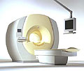
From Philips Medical Systems;
the Intera-family offers with this member a wide range of possibilities, efficiency and a ergonomic and intuitive serving-platform. Also available as Intera CV for cardiac and Intera I/T for interventional MR procedures.
The scanners are also equipped with SENSE technology, which is essential for high-quality contrast enhanced magnetic resonance angiography, interactive cardiac MR and diffusion tensor imaging ( DTI) fiber tracking.
The increased accuracy and clarity of MR scans obtained with this technology allow for faster and more accurate diagnosis of potential problems like patient friendliness and expands the breadth of applications including cardiology, oncology and interventional MR.
Device Information and Specification
CLINICAL APPLICATION
Whole body
CONFIGURATION
Short bore compact
Standard: head, body, C1, C3; Optional: Small joint, flex-E, flex-R, endocavitary (L and S), dual TMJ, knee, neck, T/L spine, breast; Optional phased array: Spine, pediatric, 3rd party connector; Optional SENSE coils: Flex-S-M-L, flex body, flex cardiac
SE, Modified-SE ( TSE), IR (T1, T2, PD), STIR, FLAIR, SPIR, FFE, T1-FFE, T2-FFE, Balanced FFE, TFE, Balanced TFE, Dynamic, Keyhole, 3D, Multi Chunk 3D, Multi Stack 3D, K Space Shutter, MTC, TSE, Dual IR, DRIVE, EPI, Cine, 2DMSS, DAVE, Mixed Mode; Angiography: PCA, MCA, Inflow MRA, CE
TR
2.9 (Omni), 1.6 (Power), 1.6 (Master/Expl) msec
TE
1.0 (Omni), 0.7 (Power), 0.5 (Master/Expl) msec
RapidView Recon. greater than 500 @ 256 Matrix
0.1 mm(Omni), 0.05 mm (Pwr/Mstr/Expl)
128 x 128, 256 x 256,512 x 512,1024 x 1024 (64 for BOLD img.)
Variable in 1% increments
Lum.: 120 cd/m2; contrast: 150:1
Variable (op. param. depend.)
POWER REQUIREMENTS
380/400 V
| |  | |
• View the DATABASE results for 'Intera 1.5T™' (2).
| | | | |
|  |  | Searchterm 'Range' was also found in the following services: | | | | |
|  |  |
| |
|
Isomers are molecules with the same chemical formula and often with the same kinds of bonds between atoms, but in which the atoms are ar ranged differently.
See also Isotope. | |  | |
• View the DATABASE results for 'Isomer' (3).
| | | | |
|  | |  |  |  |
| |
|
| |  | |
• View the DATABASE results for 'Larmor Equation' (6).
| | | | |  Further Reading: Further Reading: | News & More:
|
|
| |
|  | |  |  |
|  | |
|  | | |
|
| |
 | Look
Ups |
| |