 | Info
Sheets |
| | | | | | | | | | | | | | | | | | | | | | | | |
 | Out-
side |
| | | | |
|
| | | | | |  | Searchterm 'Range' was also found in the following services: | | | | |
|  |  |
| |
|
An array coil combines the advantages of smaller coils (high SNR) with those of larger coils (large measurement field).
This type of RF coil is composed of separate multiple smaller coils, which can be used individually ( switchable coil) or combined.
When used simultaneously, the elements can either be:
•
coupled array coils - electrically coupled to each other through common transmission lines or mutual inductance
•
isolated array coils - electrically isolated from each other with separate transmission lines and receivers and minimum effective
mutual inductance, and with the signals from each transmission line processed independently or at different frequencies
•
phased array coils - multiple small coils arranged to efficiently cover a specific anatomic region and obtain high-resolution, high-SNR images of a larger volume. The data from the individual coils is integrated by special software to produce the high-resolution images.
See also the related poll result: ' 3rd party coils are better than the original manufacturer coils'
| | | |  | | | | | | | | |  Further Reading: Further Reading: | News & More:
|
|
| |
|  | |  |  |  |
| |
|
A RF coil, often a transmit receive coil with a number of wires running along the z-direction, ar ranged to give a cosine current variation around the circumference of the coil, which looks like a bird cage.
The bird cage coil works on a different principle to conventionally tuned local and surround coils in that it behaves like a tuned transmission line with one complete cycle of standing wave around the circumference. The frequency supply is generated by an oscillator, which is modulated to form a shaped pulse by a product detector controlled by the waveform generator. The signal must be amplified to 1000's of watts. This can be done using either solid state electronics, valves or a combination of both.
The bird cage coil design provides the best field homogeneity of all RF imaging coils.
One advantage is that it is simple to produce an exceedingly uniform B1 radio frequency field over most of the coil's volume, with the result of images with a high degree of uniformity.
A second advantage is that nodes with zero voltage occur 90° away from the driven part of the coil, thus facilitating the introduction of a second signal in quadrature, which produces a circularly polarized radio frequency field.
This type of volume coil is used for brain (head) MRI, or MR imaging of joints, such as the wrist or knees.
See also the related poll result: ' 3rd party coils are better than the original manufacturer coils' | | | |  | |
• View the DATABASE results for 'Bird Cage Coil' (4).
| | | | |  Further Reading: Further Reading: | | Basics:
|
|
News & More:
| |
| |
|  | |  |  |  |
| |
|

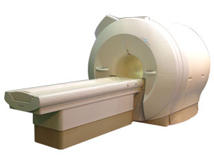
'Next generation MRI system 1.5T CHORUS developed by ISOL Technology is optimized for both clinical diagnostic imaging and for research development.
CHORUS offers the complete range of feature oriented advanced imaging techniques- for both clinical routine and research. The compact short bore magnet, the patient friendly design and the gradient technology make the innovation to new degree of perfection in magnetic resonance.'
Device Information and Specification
CLINICAL APPLICATION
Whole body
Spin Echo, Gradient Echo, Fast Spin Echo,
Inversion Recovery ( STIR, Fluid Attenuated Inversion Recovery), FLASH, FISP, PSIF, Turbo Flash ( MPRAGE ),TOF MR Angiography, Standard echo planar imaging package (SE-EPI, GE-EPI), Optional:
Advanced P.A. Imaging Package (up to 4 ch.), Advanced echo planar imaging package,
Single Shot and Diffusion Weighted EPI, IR/FLAIR EPI
STRENGTH
20 mT/m (Upto 27 mT/m)
| |  | |
• View the DATABASE results for 'CHORUS 1.5T™' (2).
| | | | |
|  |  | Searchterm 'Range' was also found in the following services: | | | | |
|  |  |
| |
|
Cervical spine MRI is a suitable tool in the assessment of all cervical spine (vertebrae C1 - C7) segments (computed tomography (CT) images may be unsatisfactory close to the thoracic spine due to shoulder artifacts). The cervical spine is particularly susceptible to degenerative problems caused by the complex anatomy and its large range of motion.
Advantages of magnetic resonance imaging MRI are the high soft tissue contrast (particularly important in diagnostics of the spinal cord), the ability to display the entire spine in sagittal views and the capacity of 3D visualization. Magnetic resonance myelography is a useful supplement to conventional MRI examinations in the investigation of cervical stenosis. Myelographic sequences result in MR images with high contrast that are similar in appearance to conventional myelograms. Additionally, open MRI studies provide the possibility of weight-bearing MRI scan to evaluate structural positional and kinetic changes of the cervical spine. Indications of cervical spine MRI scans include the assessment of soft disc herniations, suspicion of disc hernia recurrence after operation, cervical spondylosis, osteophytes, joint arthrosis, spinal canal lesions (tumors, multiple sclerosis, etc.), bone diseases (infection, inflammation, tumoral infiltration) and paravertebral spaces.
State-of-the-art phased array spine coils and high performance MRI machines provide high image quality and short scan time. Imaging protocols for the cervical spine includes sagittal T1 weighted and T2 weighted sequences with 3-4 mm slice thickness and axial slices; usually contiguous from C2 through T1. Additionally, T2 fat suppressed and T1 post contrast images are often useful in spine imaging. See also Lumbar Spine MRI.
| |  | |
• View the DATABASE results for 'Cervical Spine MRI' (2).
| | |
• View the NEWS results for 'Cervical Spine MRI' (1).
| | | | |  Further Reading: Further Reading: | News & More:
|
|
| |
|  | |  |  |  |
| |
|
A series of rapidly recorded multiple images taken at sequential cycles of time and displayed on a monitor in a dynamic movie display format. This technique can be used to show true range of motion studies of joints and parts of the spine. | | | |  | |
• View the DATABASE results for 'Cine' (57).
| | |
• View the NEWS results for 'Cine' (31).
| | | | |  Further Reading: Further Reading: | News & More:
|
|
| |
|  | |  |  |
|  | | |
|
| |
 | Look
Ups |
| |