 | Info
Sheets |
| | | | | | | | | | | | | | | | | | | | | | | | |
 | Out-
side |
| | | | |
|
| | | | |
Result : Searchterm 'MEDIC Technique' found in 1 term [ ] and 0 definition [ ] and 0 definition [ ], (+ 19 Boolean[ ], (+ 19 Boolean[ ] results ] results
| | previous 11 - 15 (of 20) nextResult Pages : [1] [2 3 4] |  | |  | Searchterm 'MEDIC Technique' was also found in the following services: | | | | |
| |  |
| |
|
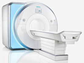
From Siemens Medical Systems;
Received FDA clearance in 2010.
MAGNETOM Skyra is a top-of-the-line, patient friendly wide bore 3 Tesla MRI system.
The system is equipped with the Tim 4G and Dot system (Total imaging matrix and Day optimizing throughput), to enhance both productivity and image quality with the complete range of advanced applications for clinical routine and research. Tim 4G features lighter, trimmer MRI coils that take up less space inside the magnet but deliver a high coil element density with increased signal to noise ratio and the possibility to use high iPAT factors.
Device Information and Specification
CLINICAL APPLICATION
Whole Body
Head, spine, torso/ body coil, neurovascular, cardiac, neck, shoulder, knee, wrist, foot//ankle and multi-purpose flex coils. Peripheral vascular, breast, shoulder.
CHANNELS (min. / max. configuration)
48, 64, 128
Chemical shift imaging, single voxel spectroscopy
MINIMUM TE
3D T1 spoiled GRE: 0.22 (256 matrix), Ultra-short TE
At isocenter: L-R 70 cm, A-P (with table) 55 cm
MAGNET WEIGHT (gantry included)
5768 kg
DIMENSION H*W*D (gantry included)
173 x 231 x 219 cm
COOLING SYSTEM
Water; single cryogen, 2 stage refrigeration
3 linear with 20 coils, 5 nonlinear 2nd-order
POWER REQUIREMENTS
380 / 400 / 420 / 440 / 460 / 480 V, 3-phase + ground; 110 kVA
| | | | | |
| | | | | |
| |
|
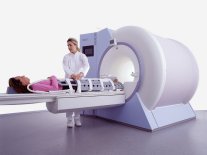
From Siemens Medical Systems;
while older navigator techniques take up to 40 minutes to create, the high performance of the MAGNETOM Sonata system enables 'complete examinations in less than 15 minutes'. It creates a new standard of diagnostic confidence and moves Cardiac MR from the research setting into routine clinical practice.
Device Information and Specification CLINICAL APPLICATION Whole body Body, head, spine, knee, neck, TMJ, extremity, head, breast, shoulder, others GRE, IR, FIR, STIR, TrueIR/FISP, FSE, FLAIR, MT, SS-FSE, MT-SE, MTC, MSE, EPI, 3D DESS//CISS/PSIF, GMR IMAGING MODES Single, multislice, volume study, multi angle, multi oblique178 images/sec at 256 x 256 at 100% FOV1024 x 1024 full screen display 4050kg, 5500kg in operation POWER REQUIREMENTS 380/400/420/440/480 V Passive, act.; 1st order std./2nd opt. | | | |
• View the DATABASE results for 'MAGNETOM Sonata™' (2).
| | | | |
| | | | | |
| |
|
( MRA) Magnetic resonance angiography is a medical imaging technique to visualize blood filled structures, including arteries, veins and the heart chambers. This MRI technique creates soft tissue contrast between blood vessels and surrounding tissues primarily created by flow, rather than displaying the vessel lumen. There are bright blood and black blood MRA techniques, named according to the appearance of the blood vessels. With this different MRA techniques both, the blood flow and the condition of the blood vessel walls can be seen. Flow effects in MRI can produce a range of artifacts. MRA takes advantage of these artifacts to create predictable image contrast due to the nature of flow.
Technical parameters of the MRA sequence greatly affect the sensitivity of the images to flow with different velocities or directions, turbulent flow and vessel size.
This are the three main types of MRA:
All angiographic techniques differentially enhance vascular MR signal. The names of the bright blood techniques TOF and PCA reflect the physical properties of flowing blood that were exploited to make the vessels appear bright. Contrast enhanced magnetic resonance angiography creates the angiographic effect by using an intravenously administered MR contrast agent to selectively shorten the T1 of blood and thereby cause the vessels to appear bright on T1 weighted images.
MRA images optimally display areas of constant blood flow-velocity, but there are many situations where the flow within a voxel has non-uniform speed or direction. In a diseased vessel these patterns are even more complex. Similar loss of streamline flow occurs at all vessel junctions and stenoses, and in regions of mural thrombosis. It results in a loss of signal, due to the loss of phase coherence between spins in the voxel.
This signal loss, usually only noticeable distal to a stenosis, used to be an obvious characteristic of MRA images. It is minimized by using small voxels and the shortest possible TE. Signal loss from disorganized flow is most noticeable in TOF imaging but also affects the PCA images.
Indications to perform a magnetic resonance angiography ( MRA):
•
Detection of aneurysms and dissections
•
Evaluation of the vessel anatomy, including variants
•
Blockage by a blood clot or stenosis of the blood vessel caused by plaques (the buildup of fat and calcium deposits)
Conventional angiography or computerized tomography angiography (CT angiography) may be needed after MRA if a problem (such as an aneurysm) is present or if surgery is being considered.
See also Magnetic Resonance Imaging MRI. | | | | | | | | | | |
• View the DATABASE results for 'Magnetic Resonance Angiography MRA' (3).
| | |
• View the NEWS results for 'Magnetic Resonance Angiography MRA' (10).
| | | | |  Further Reading: Further Reading: | | Basics:
|
|
News & More:
| |
| |
| | | Searchterm 'MEDIC Technique' was also found in the following services: | | | | |
| | |
| |
|
(DTI) Diffusion tensor imaging is the more sophisticated form of DWI, which allows for the determination of directionality as well as the magnitude of water diffusion. This kind of MR imaging can estimates damage to nerve fibers that connect the area of the brain affected by the stroke to brain regions that are distant from it, and can be used to determine the effectiveness of stroke prevention medications.
DTI (FiberTrak) enables to visualize white matter fibers in the brain and can map ( trace image) subtle changes in the white matter associated with diseases such as multiple sclerosis and epilepsy, as well as assessing diseases where the brain's wiring is abnormal, such as schizophrenia.
The fractional anisotropy (FA) gives information about the shape of
the diffusion tensor at each voxel. The FA is based on the normalized
variance of the eigenvalues. The fractional anisotropy reflects differences between an isotropic diffusion and a linear diffusion. The FA range is between 0 and 1 (0 = isotropic diffusion, 1 = highly directional).
The development of new imaging methods and some useful analysis techniques, such as 3-dimensional anisotropy contrast (3DAC) and spatial tracking of the diffusion tensor tractography (DTT), are currently under study. | | | |
• View the DATABASE results for 'Diffusion Tensor Imaging' (9).
| | |
• View the NEWS results for 'Diffusion Tensor Imaging' (2).
| | | | | | Further Reading: | | Basics:
|
|
News & More:
| |
| |
| | | | | |
| |
|
(ESR) Electron spin resonance is a spectroscopic technique to identify paramagnetic substances. This magnetic resonance phenomenon investigates the nature of the bonding within molecules by identifying unpaired electrons, e.g. in free radicals and their interaction with their immediate surroundings. The Larmor frequency are much higher than corresponding NMR frequencies in the same static magnetic field.
Nuclei with an odd number of neutrons and/or protons, because of their spin, react like tiny magnets and can be lined up in an applied magnetic field. Energy applied by alternating radio frequency radiation is absorbed when its frequency coincides with that of precession of the electron magnets. The spectrum of radiation absorbed as the field changes gives information valuable in chemistry, biology, and medicine since over 50 years. | | | |
• View the DATABASE results for 'Electron Spin Resonance' (2).
| | |
• View the NEWS results for 'Electron Spin Resonance' (1).
| | | | | | Further Reading: | Basics:
|
|
News & More:
| |
| |
| | | | |
| | | |
|
| |
 | Look
Ups |
| |


