 | Info
Sheets |
| | | | | | | | | | | | | | | | | | | | | | | | |
 | Out-
side |
| | | | |
|
| | | | |
Result : Searchterm 'Array' found in 8 terms [ ] and 53 definitions [ ] and 53 definitions [ ] ]
| | previous 46 - 50 (of 61) nextResult Pages : [1 2] [3 4 5 6 7 8 9 10 11 12 13] |  | |  | Searchterm 'Array' was also found in the following services: | | | | |
| |  |
| |
|
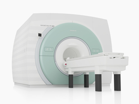
Device Information and Specification
CLINICAL APPLICATION
Whole body
CONFIGURATION
Short bore compact
Standard: Head, body, cardiac, optional phased array: Spine, pediatric, 3rd party connector; Optional SENSE? coils for all applications
SE, Modified-SE, IR (T1, T2, PD), STIR, FLAIR, SPIR, FFE, T1-FFE, T2-FFE, Balanced FFE, TFE, Balanced TFE, Dynamic, Keyhole, 3D, Multi Chunk 3D, Multi Stack 3D, K Space Shutter, MTC, TSE, Dual IR, DRIVE, EPI, Cine, 2DMSS, DAVE, Mixed Mode; Angiography: Inflow MRA, TONE, PCA, CE MRA
128 x 128, 256 x 256,512 x 512,1024 x 1024 (64 for Bold img)
Variable in 1% increments
Lum.: 120 cd/m2; contrast: 150:1
Variable (op. param. depend.)
POWER REQUIREMENTS
380/400 V
| | | | | |  Further Reading: Further Reading: | News & More:
|
|
| |
| | | | | |
| |
|
| | | |
• View the DATABASE results for 'Liver Acquisition with Volume Acquisition' (3).
| | | | | | Further Reading: | Basics:
|
|
| |
| | | | | |
| |
|
(LUT) A lookup table is a data structure, usually an array. In MRI for example the intensity values are mapped to the related image pixels. | | | |
• View the DATABASE results for 'Lookup Table' (2).
| | | | |
| | | Searchterm 'Array' was also found in the following services: | | | | |
| | |
| |
|
MRI of the lumbar spine, with its multiplanar 3 dimensional imaging capability, is currently the preferred modality for establishing a diagnosis. MRI scans and magnetic resonance myelography have many advantages compared with computed tomography and/or X-ray myelography in evaluating the lumbar spine. MR imaging scans large areas of the spine without ionizing radiation, is noninvasive, not affected by bone artifacts, provides vascular imaging capability, and makes use of safer contrast agents ( gadolinium chelate).
Due to the high level of tissue contrast resolution, nerves and discs are clearly visible. MRI is excellent for detecting degenerative disease in the spine. Lumbar spine MRI accurately shows disc disease (prolapsed disc or slipped disc), the level at which disc disease occurs, and if a disc is compressing spinal nerves. Lumbar spine MRI depicts soft tissues, including the cauda equina, spinal cord, ligaments, epidural fat, subarachnoid space, and intervertebral discs. Loss of epidural fat on T1 weighted images, loss of cerebrospinal fluid signal around the dural sac on T2 weighted images and degenerative disc disease are common features of lumbar stenosis.
Common indications for MRI of the lumbar spine:
•
Neurologic deficits, evidence of radiculopathy, acute spinal cord compression (e.g., sudden bowel/bladder disturbance)
•
Suspected systemic disorders (primary tumors, drop metastases, osteomyelitis)
•
Postoperative evaluation of lumbar spine: disk vs. scar
•
Localized back pain with no radiculopathy (leg pain)
Lumbar spine imaging requires a special spine coil. often used whole spine array coils have the advantage that patients do not need other positioning if also upper parts of the spine should be scanned. Sagittal T1 and T2 weighted FSE sequences are the standard views. With multi angle oblique techniques individually oriented transverse images of each intervertebral disc at different angles can be obtained.
See also the related poll result: ' MRI will have replaced 50% of x-ray exams by' | | | | | |
• View the DATABASE results for 'Lumbar Spine MRI' (6).
| | | | | | Further Reading: | | Basics:
|
|
News & More:
| |
| |
| | | | | |
| |
|
From Siemens Medical Systems;
The MAGNETOM 7T is designed as an open research platform. 7T MRI provides anatomical detail at the submillimiter scale, enhanced contrast mechanisms, outstanding spectroscopy performance, ultra-high resolution functional imaging ( fMRI), multinuclear whole-body MRI and functional information.
This ultra high field (UHF) MRI device is a research system and not cleared, approved or licensed in any jurisdiction for patient examinations.
Device Information and Specification
CLINICAL APPLICATION
Whole body
High-performance, ultra high field coils available. Integration and support for coil developments.
CHANNELS (min. / max. configuration)
32, optional 8 channels TX array
40 x 40 x 30 cm³ less than 8% nonlinearity
MAGNET WEIGHT (gantry included)
35017 kg
DIMENSION H*W*D (gantry included)
320 x 240 x 317,5 cm
MAX. AMPLITUDE
up to 70 mT/m
Up to 3rd order shim coils, user configurable B0 shim ? B0 maps and ROI definition
POWER REQUIREMENTS
2000 Volts, 650A
| | | | | | | Further Reading: | | Basics:
|
|
News & More:
| |
| |
| | | | |
| | | |
|
| |
 | Look
Ups |
| |


