 | Info
Sheets |
| | | | | | | | | | | | | | | | | | | | | | | | |
 | Out-
side |
| | | | |
|
| | | | | |  | Searchterm 'Arc' was also found in the following services: | | | | |
|  |  |
| |
|

From Philips Medical Systems;
the Intera 3 T high field system, the first with a compact magnet, which is built on the same platform as the 1.5 T, is targeted to high-end neurological, orthopedic and cardiovascular imaging applications with maximum patient comfort and acceptance without compromising image quality and clinical performance. Useable for clinical routine and rese arch.
The Intera systems offer diffusion tensor imaging ( DTI) fiber tracking that measures movement of water in the brain and can therefore detect areas of the brain where normal movement of water is disrupted.
Device Information and Specification
CLINICAL APPLICATION
Whole body
CONFIGURATION
Short bore compact
Standard: head, body, C1, C3; Optional: Small joint, flex-E, flex-R, endocavitary (L and S), dual TMJ, knee, neck, T/L spine, breast; Optional phased array: spine;; Optional SENSE coils: Flex body, flex cardiac, neuro-vascular, head
SE, Modified-SE, IR (T1, T2, PD), STIR, FLAIR, SPIR, FFE, T1-FFE, T2-FFE, Balanced FFE, TFE, Balanced TFE, Dynamic, Keyhole, 3D, Multi Chunk 3D, Multi Stack 3D, K Space Shutter, MTC, TSE, Dual IR, DRIVE, EPI, Cine, 2DMSS, DAVE, Mixed Mode; Angiography: Inflow MRA, TONE, PCA, CE MRA
TR
Min. 1.6 (Master) msec
TE
Min. 0.5 (Master) msec
RapidView Recon. greater than 500 @ 256 Matrix
0.1 mm (Omni), 0.05 mm (Power)
128 x 128, 256 x 256,512 x 512,1024 x 1024 (64 for Bold img)
Variable in 1% increments
Lum.: 120 cd/m2; contrast: 150:1
Variable (op. param. depend.)
POWER REQUIREMENTS
380/400 V
STRENGTH
30 (Master) mT/m
| | | | | |
| | | Searchterm 'Arc' was also found in the following services: | | | | |
| | |
| |
|
Device Information and Specification
CLINICAL APPLICATION
Whole body
CONFIGURATION
Short bore compact
Standard: Head, body, cardiac, optional phased array: Spine, pediatric, 3rd party connector; Optional SENSE? coils for all applications
SE, Modified-SE, IR (T1, T2, PD), STIR, FLAIR, SPIR, FFE, T1-FFE, T2-FFE, Balanced FFE, TFE, Balanced TFE, Dynamic, Keyhole, 3D, Multi Chunk 3D, Multi Stack 3D, K Space Shutter, MTC, TSE, Dual IR, DRIVE, EPI, Cine, 2DMSS, DAVE, Mixed Mode; Angiography: Inflow MRA, TONE, PCA, CE MRA
128 x 128, 256 x 256,512 x 512,1024 x 1024 (64 for Bold img)
Variable in 1% increments
Lum.: 120 cd/m2; contrast: 150:1
Variable (op. param. depend.)
POWER REQUIREMENTS
380/400 V
| | | |
• View the DATABASE results for 'Intera Achieva CV™' (2).
| | | | |  Further Reading: Further Reading: | News & More:
|
|
| |
| | | | | |
| |
|
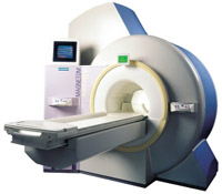
From Siemens Medical Systems;
the 3 T MAGNETOM Allegra is a dedicated MR headscanner, perfect as a rese arch system in cognitive and neuroscience with MRS and fMRI. MAGNETOM Allegra is a full member of the MAGNETOM product family. It uses many common components, i.e. electronics, computer system, software and pulse sequence concepts.
Device Information and Specification
GRE, IR, FIR, STIR, TrueIR/FISP, FSE, FLAIR, MT, SS-FSE, MT-SE, MTC, MSE, EPI, GMR, fat/water sat./exc.
IMAGING MODES
Single, multislice, volume study, multi angle, multi oblique
178 images/sec at 256 x 256 at 100% FOV
1024 x 1024 full screen display
MAGNET WEIGHT (gantry included)
5500 kg
DIMENSION H*W*D (gantry included)
220 x 220 x 147 cm
POWER REQUIREMENTS
380/400/420/440/480 V
Passive, act.; 1st order std./2nd opt.
| | | |
• View the DATABASE results for 'MAGNETOM Allegra™' (2).
| | | | |
| | | Searchterm 'Arc' was also found in the following services: | | | | |
| | |
| |
|
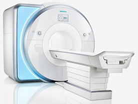
From Siemens Medical Systems;
Received FDA clearance in 2013.
The MAGNETOM Prisma is the 3T PowerPack for exploration that offers most demanding clinical and rese arch challenges of today and the future. The latest parallel transmit technology, TimTX TrueShape, enables zooming into specific body regions for enhanced image quality. Furthermore, the Tim 4G integrated coil technology offers remarkable imaging flexibility and supports complex
examinations across the whole body.
Onsite upgrades to the MAGNETOM Prisma for customers who have already installed the 3 Tesla MAGNETOM Trio are possible.
Device Information and Specification
CLINICAL APPLICATION
Whole Body
CONFIGURATION
Ultra-short bore
Head, spine, torso/ body coil, neurovascular, cardiac, neck, shoulder, knee, wrist, foot//ankle and multi-purpose flex coils. Peripheral vascular, breast, shoulder.
CHANNELS (min. / max. configuration)
64, 128
MAGNET WEIGHT (gantry included)
13000 kg
DIMENSION H*W*D (gantry included)
173 x 230 x 222 cm
Passive, active; first order,
second order
POWER REQUIREMENTS
380 / 400 / 420 / 440 / 460 / 480 V, 3-phase + ground;
| | | | | |
| | | Searchterm 'Arc' was also found in the following services: | | | | |
| | |
| |
|
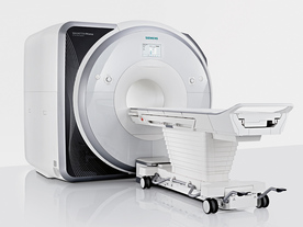
From Siemens Medical Systems;
Received FDA clearance in 2010.
MAGNETOM Skyra is a top-of-the-line, patient friendly wide bore 3 Tesla MRI system.
The system is equipped with the Tim 4G and Dot system (Total imaging matrix and Day optimizing throughput), to enhance both productivity and image quality with the complete range of advanced applications for clinical routine and rese arch. Tim 4G features lighter, trimmer MRI coils that take up less space inside the magnet but deliver a high coil element density with increased signal to noise ratio and the possibility to use high iPAT factors.
Device Information and Specification
CLINICAL APPLICATION
Whole Body
Head, spine, torso/ body coil, neurovascular, cardiac, neck, shoulder, knee, wrist, foot//ankle and multi-purpose flex coils. Peripheral vascular, breast, shoulder.
CHANNELS (min. / max. configuration)
48, 64, 128
Chemical shift imaging, single voxel spectroscopy
MINIMUM TE
3D T1 spoiled GRE: 0.22 (256 matrix), Ultra-short TE
At isocenter: L-R 70 cm, A-P (with table) 55 cm
MAGNET WEIGHT (gantry included)
5768 kg
DIMENSION H*W*D (gantry included)
173 x 231 x 219 cm
COOLING SYSTEM
Water; single cryogen, 2 stage refrigeration
3 linear with 20 coils, 5 nonlinear 2nd-order
POWER REQUIREMENTS
380 / 400 / 420 / 440 / 460 / 480 V, 3-phase + ground; 110 kVA
| | | | | |
| | | | |
| | |
| | | |
|
| |
 | Look
Ups |
| |


