 | Info
Sheets |
| | | | | | | | | | | | | | | | | | | | | | | | |
 | Out-
side |
| | | | |
|
| | | | |
Result : Searchterm 'panorama' found in 9 terms [ ] and 1 definition [ ] and 1 definition [ ] ]
| | previous 6 - 10 (of 10) Result Pages : [1 2] |  | |  | Searchterm 'panorama' was also found in the following services: | | | | |
| |  |
| |
|

From Philips Medical Systems;
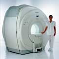
the Intera 3 T high field system, the first with a compact magnet, which is built on the same platform as the 1.5 T, is targeted to high-end neurological, orthopedic and cardiovascular imaging applications with maximum patient comfort and acceptance without compromising image quality and clinical performance. Useable for clinical routine and research.
The Intera systems offer diffusion tensor imaging ( DTI) fiber tracking that measures movement of water in the brain and can therefore detect areas of the brain where normal movement of water is disrupted.
Device Information and Specification
CLINICAL APPLICATION
Whole body
CONFIGURATION
Short bore compact
Standard: head, body, C1, C3; Optional: Small joint, flex-E, flex-R, endocavitary (L and S), dual TMJ, knee, neck, T/L spine, breast; Optional phased array: spine;; Optional SENSE coils: Flex body, flex cardiac, neuro-vascular, head
SE, Modified-SE, IR (T1, T2, PD), STIR, FLAIR, SPIR, FFE, T1-FFE, T2-FFE, Balanced FFE, TFE, Balanced TFE, Dynamic, Keyhole, 3D, Multi Chunk 3D, Multi Stack 3D, K Space Shutter, MTC, TSE, Dual IR, DRIVE, EPI, Cine, 2DMSS, DAVE, Mixed Mode; Angiography: Inflow MRA, TONE, PCA, CE MRA
TR
Min. 1.6 (Master) msec
TE
Min. 0.5 (Master) msec
RapidView Recon. greater than 500 @ 256 Matrix
0.1 mm (Omni), 0.05 mm (Power)
128 x 128, 256 x 256,512 x 512,1024 x 1024 (64 for Bold img)
Variable in 1% increments
Lum.: 120 cd/m2; contrast: 150:1
Variable (op. param. depend.)
POWER REQUIREMENTS
380/400 V
STRENGTH
30 (Master) mT/m
| | | | | |
| | | | | |
| |
|
From Philips Medical Systems;
Philips Infinion 1.5 T is designed to maximize the efficiency and quality of patient care. Developed with the patient in mind, the Infinion is the shortest and most open 1.5T scanner available. The unique 'ultra short' 1.4 m magnet assures patient comfort and acceptance without compromising image quality and clinical performance.
Device Information and Specification
CLINICAL APPLICATION
Whole body
CONFIGURATION
Ultra short bore
Head, head / neck, integrated C-spine, L/T spine array, small large GP coils, body flex array, torso pelvis array, breast array, endocavitary, shoulder array, lower extremity, hand / wrist, cardiac, PV array
SE, TSE, SS TSE, EPI, IR, STIR, FLAIR, FFE, TFE, T1 TFE, T2 TFE, Presat, Fatsat, MTC, Diff-opt., Angiography: PCA, MCA, TOF
IMAGING MODES
Single slice, single volume, multi slice, multi volume
80 images/sec std.; up to320 opt.@256
H*W*D
233 (lead fitted) x 198 x 140 cm
POWER REQUIREMENTS
400/480 V
COOLING SYSTEM TYPE
Closed loop, chilled water
| | | |
• View the DATABASE results for 'Infinion 1.5T™' (2).
| | | | |
| | | | | |
| |
|
Multiple independent array coil systems can be connected simultaneously and up to 4 coil elements can be combined for a measurement. | | | | | |
| | | Searchterm 'panorama' was also found in the following services: | | | | |
| | |
| |
|
Device Information and Specification CLINICAL APPLICATION Whole body SE, FE, IR, STIR, FFE, DEFFE, DESE, TSE, DETSE, Single shot SE, DRIVE, Balanced FFE, MRCP, Fluid Attenuated Inversion Recovery, Turbo FLAIR, IR-TSE, T1-STIR TSE, T2-STIR TSE, Diffusion Imaging, 3D SE, 3D FFE, Contrast Perfusion Analysis, MTC;; Angiography: CE-ANGIO, MRA 2D, 3D TOFOpen x 47 cm x infinite (side-first patient entry) POWER REQUIREMENTS 400/480 V | | | |
• View the DATABASE results for 'Panorama 0.6T™' (2).
| | | | |
| | | | |  |
| |
|

Philips Medical System is the diagnostics business of Royal Philips Electronics of the Netherlands, one of the world's biggest electronics companies and Europe's largest. Philips is quoted on the NYSE (symbol: PHG), London, Frankfurt, Amsterdam and other stock exchanges.
On October 19, 2001, Philips Medical Systems completed a 3-year acquisition strategy through its purchase of Marconi Medical Systems. Marconi Medical Systems offered leading multislice CT, MRI, and Nuclear Gamma Camera systems to medical institutions around the world. As well as new 3.0T developments, Philips is also in collaboration with researchers at the University of Nottingham, with the intention of developing an ultrahigh field strength clinical 7.0T whole body MR system.
MRI Scanners:
0.23T to 1.0T:
1.5T:
3.0T to 7.0T:
Hybrid Scanners:
Contact Information MAIL Philips Medical Systems
3000 Minuteman Road
Andover, MA 01810-1099
USA | | | |
• View the DATABASE results for 'Philips Medical Systems' (14).
| | |
• View the NEWS results for 'Philips Medical Systems' (12).
| | | | |  Further Reading: Further Reading: | News & More:
|
|
| |
| | | | |
| | |
| | | |
|
| |
 | Look
Ups |
| |


