 | Info
Sheets |
| | | | | | | | | | | | | | | | | | | | | | | | |
 | Out-
side |
| | | | |
|
| | | | |
Result : Searchterm 'Time Inversion' found in 1 term [ ] and 0 definition [ ] and 0 definition [ ], (+ 19 Boolean[ ], (+ 19 Boolean[ ] results ] results
| | previous 6 - 10 (of 20) nextResult Pages : [1] [2 3 4] |  | | | | |  |
| |
|
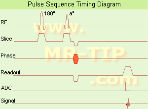
In simple ultrafast GRE imaging, TR and TE are so short, that tissues have a poor imaging signal and - more importantly - poor contrast except when contrast media enhanced ( contrast enhanced angiography). Therefore, the magnetization is 'prepared' during the preparation module, most frequently by an initial 180° inversion pulse.
In the pulse sequence timing diagram, the basic ultrafast gradient echo sequence is illustrated. The 180° inversion pulse is executed one time (to the left of the vertical line), the right side represents the data collection period and is often repeated depending on the acquisition parameters.
See also Pulse Sequence Timing Diagram, there you will find a description of the components.
Ultrafast GRE sequences have a short TR,TE, a low flip angle and TR is so short that image acquisition lasts less than 1 second and typically less than 500 ms. Common TR: 3-5 msec, TE: 2 msec, and the flip angle is about 5°.
Such sequences are often labeled with the prefix 'Turbo' like TurboFLASH, TurboFFE and TurboGRASS.
This allows one to center the subsequent ultrafast GRE data acquisition around the inversion time TI, where one of the tissues of interest has very little signal as its z-magnetization is passing through zero.
Unlike a standard inversion recovery (IR) sequence, all lines or a substantial segment of k-space image lines are acquired after a single inversion pulse, which can then together be considered as readout module. The readout module may use a variable flip angle approach, or the data acquisition may be divided into multiple segments (shots). The latter is useful particularly in cardiac imaging where acquiring all lines in a single segment may take too long relative to the cardiac cycle to provide adequate temporal resolution.
If multiple lines are acquired after a single pulse, the pulse sequence is a type of gradient echo echo planar imaging (EPI) pulse sequence. See also Magnetization Prepared Rapid Gradient Echo ( MPRAGE) and Turbo Field Echo ( TFE). | | | | | | | | | | |
| | | | | |
| |
|

'Next generation MRI system 1.5T CHORUS developed by ISOL Technology is optimized for both clinical diagnostic imaging and for research development.
CHORUS offers the complete range of feature oriented advanced imaging techniques- for both clinical routine and research. The compact short bore magnet, the patient friendly design and the gradient technology make the innovation to new degree of perfection in magnetic resonance.'
Device Information and Specification
CLINICAL APPLICATION
Whole body
Spin Echo, Gradient Echo, Fast Spin Echo,
Inversion Recovery ( STIR, Fluid Attenuated Inversion Recovery), FLASH, FISP, PSIF, Turbo Flash ( MPRAGE ),TOF MR Angiography, Standard echo planar imaging package (SE-EPI, GE-EPI), Optional:
Advanced P.A. Imaging Package (up to 4 ch.), Advanced echo planar imaging package,
Single Shot and Diffusion Weighted EPI, IR/FLAIR EPI
STRENGTH
20 mT/m (Upto 27 mT/m)
| | | |
• View the DATABASE results for 'CHORUS 1.5T™' (2).
| | | | |
| | | | | |
| |
|

'MRI system is not an expensive equipment anymore.
ENCORE developed by ISOL Technology is a low cost MRI system with the advantages like of the 1.0T MRI scanner. Developed specially for the overseas market, the ENCORE is gaining popularity in the domestic market by medium sized hospitals.
Due to the optimum RF and Gradient application technology. ENCORE enables to obtain high resolution imaging and 2D/3D Angio images which was only possible in high field MR systems.'
- Less consumption of the helium gas due to the ultra-lightweight magnet specially designed and manufactured for ISOL.
- Cost efficiency MR system due to air cooling type (equivalent to permanent magnetic).
- Patient processing speed of less than 20 minutes.'
Device Information and Specification
CLINICAL APPLICATION
Whole body
CONFIGURATION
Short bore compact
| | | |
• View the DATABASE results for 'ENCORE 0.5T™' (2).
| | | | |
| | | | | |
| |
|
| | | |
• View the DATABASE results for 'Adiabatic Fast Passage' (3).
| | | | |  Further Reading: Further Reading: | | Basics:
|
|
News & More:
| |
| |
| | | | | |
| |
|
(CSI) Chemical shift imaging is an extension of MR spectroscopy, allowing metabolite information to be measured in an extended region and to add the chemical analysis of body tissues to the potential clinical utility of Magnetic Resonance. The spatial location is phase encoded and a spectrum is recorded at each phase encoding step to allow the spectra acquisition in a number of volumes covering the whole sample. CSI provides mapping of chemical shifts, analog to individual spectral lines or groups of lines.
Spatial resolution can be in one, two or three dimensions, but with long acquisition times od full 3D CSI. Commonly a slice-selected 2D acquisition is used. The chemical composition of each voxel is represented by spectra, or as an image in which the signal intensity depends on the concentration of an individual metabolite. Alternatively frequency-selective pulses excite only a single spectral component.
There are several methods of performing chemical shift imaging, e.g. the inversion recovery method, chemical shift selective imaging sequence, chemical shift insensitive slice selective RF pulse, the saturation method, spatial and chemical shift encoded excitation and quantitative chemical shift imaging.
See also Magnetic Resonance Spectroscopy. | | | |
• View the DATABASE results for 'Chemical Shift Imaging' (6).
| | | | | | Further Reading: | | Basics:
|
|
News & More:
| |
| |
| | | | |
| | | |
|
| |
 | Look
Ups |
| |


