| Info
Sheets |
| | | | | | | | | | | | | | | | | | | | | | | | |
 | Out-
side |
| | | | |
|
| | | | |
Result : Searchterm 'Solenoid' found in 4 terms [ ] and 11 definitions [ ] and 11 definitions [ ] ]
| | previous 6 - 10 (of 15) nextResult Pages : [1] [2 3] |  | | | | |  |
| |
|
Magnetic resonance imaging ( MRI) is based on the magnetic resonance phenomenon, and is used for medical diagnostic imaging since ca. 1977 (see also MRI History).
The first developed MRI devices were constructed as long narrow tunnels. In the meantime the magnets became shorter and wider. In addition to this short bore magnet design, open MRI machines were created. MRI machines with open design have commonly either horizontal or vertical opposite installed magnets and obtain more space and air around the patient during the MRI test.
The basic hardware components of all MRI systems are the magnet, producing a stable and very intense magnetic field, the gradient coils, creating a variable field and radio frequency (RF) coils which are used to transmit energy and to encode spatial positioning. A computer controls the MRI scanning operation and processes the information.
The range of used field strengths for medical imaging is from 0.15 to 3 T. The open MRI magnets have usually field strength in the range 0.2 Tesla to 0.35 Tesla. The higher field MRI devices are commonly solenoid with short bore superconducting magnets, which provide homogeneous fields of high stability.
There are this different types of magnets:
The majority of superconductive magnets are based on niobium-titanium (NbTi) alloys, which are very reliable and require extremely uniform fields and extreme stability over time, but require a liquid helium cryogenic system to keep the conductors at approximately 4.2 Kelvin (-268.8° Celsius). To maintain this temperature the magnet is enclosed and cooled by a cryogen containing liquid helium (sometimes also nitrogen).
The gradient coils are required to produce a linear variation in field along one direction, and to have high efficiency, low inductance and low resistance, in order to minimize the current requirements and heat deposition. A Maxwell coil usually produces linear variation in field along the z-axis; in the other two axes it is best done using a saddle coil, such as the Golay coil.
The radio frequency coils used to excite the nuclei fall into two main categories; surface coils and volume coils.
The essential element for spatial encoding, the gradient coil sub-system of the MRI scanner is responsible for the encoding of specialized contrast such as flow information, diffusion information, and modulation of magnetization for spatial tagging.
An analog to digital converter turns the nuclear magnetic resonance signal to a digital signal. The digital signal is then sent to an image processor for Fourier transformation and the image of the MRI scan is displayed on a monitor.
For Ultrasound Imaging (USI) see Ultrasound Machine at Medical-Ultrasound-Imaging.com.
See also the related poll results: ' In 2010 your scanner will probably work with a field strength of' and ' Most outages of your scanning system are caused by failure of' | | | | | | | | | | | | | | | |  Further Reading: Further Reading: | News & More:
|
 |
small-steps-can-yield-big-energy-savings-and-cut-emissions-mris
Thursday, 27 April 2023 by www.itnonline.com | | |
Portable MRI can detect brain abnormalities at bedside
Tuesday, 8 September 2020 by news.yale.edu | | |
Point-of-Care MRI Secures FDA 510(k) Clearance
Thursday, 30 April 2020 by www.diagnosticimaging.com | | |
World's First Portable MRI Cleared by FDA
Monday, 17 February 2020 by www.medgadget.com | | |
Low Power MRI Helps Image Lungs, Brings Costs Down
Thursday, 10 October 2019 by www.medgadget.com | | |
Cheap, portable scanners could transform brain imaging. But how will scientists deliver the data?
Tuesday, 16 April 2019 by www.sciencemag.org | | |
The world's strongest MRI machines are pushing human imaging to new limits
Wednesday, 31 October 2018 by www.nature.com | | |
Kyoto University and Canon reduce cost of MRI scanner to one tenth
Monday, 11 January 2016 by www.electronicsweekly.com | | |
A transportable MRI machine to speed up the diagnosis and treatment of stroke patients
Wednesday, 22 April 2015 by medicalxpress.com | | |
Portable 'battlefield MRI' comes out of the lab
Thursday, 30 April 2015 by physicsworld.com | | |
Chemists develop MRI technique for peeking inside battery-like devices
Friday, 1 August 2014 by www.eurekalert.org | | |
New devices doubles down to detect and map brain signals
Monday, 23 July 2012 by scienceblog.com |
|
| |
| | | | | |
| |
|
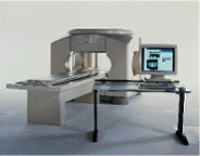
From Toshiba America Medical Systems Inc.;
FLEXART™ series is a 0.5 T superconducting MRI system that has been designed to meet the expanding role of MRI in today's clinical environment. The system utilizes innovative technologies such as digital RF, high speed actively shielded gradients and optimized RF coils which support a wide range of MRI developments.
Device Information and Specification
CLINICAL APPLICATION
Whole body
Quadrature, solenoid and multi-channel configurations
SE, FE, IR, FastSE, FastIR, FastFLAIR, Fast STIR, FastFE, FASE, Hybrid EPI, Multi Shot EPI; Angiography: 2D(gate/non-gate)/3D TOF, SORS-STC
IMAGING MODES
Single, multislice, volume study
POWER REQUIREMENTS
380/400/415/440/480 V
COOLING SYSTEM TYPE
Closed-loop water-cooled
| | | |
• View the DATABASE results for 'FLEXART™' (2).
| | | | |
| | | | | |
| |
|
A magnet is by definition an object with magnetic properties ( magnetism) that attracts iron and produces a magnetic field. It can be a permanent magnet or an electromagnet.
Permanent magnets do not rely upon outside influences to generate their field. In permanent magnets are the atoms and molecules ordered in long range. The specific electron configuration and the distance of the atoms is what lead to this long range ordering. The electrons exist in a lower energy state if they all have the same orientation. Magnetic domains can be likened to microscopic neighborhoods in which there is a strong reinforcing interaction between particles, resulting in a high degree of order. The greater the degree of ordering within and between domains, the greater the resulting field will be. Long range ordering is one of the hallmarks of a ferromagnetic material.
A current carrying conductor for example a piece of wire, produces a magnetic field that encircles the wire. An electromagnet, in its simplest form, is a wire that has been coiled into one or more loops. This coil is known as a solenoid. The more loops of wire and the greater the current, the stronger the field will be.
Superconducting magnets are a special type of electromagnets, often used in MRI machines with high field strength. | | | |
• View the DATABASE results for 'Magnet' (669).
| | |
• View the NEWS results for 'Magnet' (315).
| | | | | | Further Reading: | | Basics:
|
|
News & More:
| |
| |
| | | | | |
| |
|
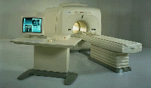
Device Information and Specification CLINICAL APPLICATION Whole body Quadrature, solenoid and multi-channel configurations SE, FE, IR, FastSE, FastIR, FastFLAIR, Fast STIR, FastFE, FASE, Hybrid EPI, Multi Shot EPI; Angiography: 2D(gate/non-gate)/3D TOF, SORS-STC IMAGING MODES Single, multislice, volume study POWER REQUIREMENTS 380/400/415/440/480 V COOLING SYSTEM TYPE Cryogenless | | | |
• View the DATABASE results for 'OPART™' (2).
| | | | |
| | | | | |
| |
|

From Philips Medical Systems;
this active shielded member of the Panorama product line combines the advantages of one 1.0 T system's with the possibilities of an open MRI system. The open design helps ease anxiety for claustrophobic patients and increased patient comfort whereby the field strength provides spectacular image quality and fast patient throughput.
Device Information and Specification CLINICAL APPLICATION Whole body Vertically opposed solenoids, head, head-neck, extremity, neck, body/ spine M-XL, shoulder, bilateral breast, wrist, TMJ, flex XS-S-M-L-XL-XXL SE, FE, IR, STIR, FFE, DEFFE, DESE, TSE, DETSE, Single shot SE, DRIVE, Balanced FFE, MRCP, FLAIR, Turbo FLAIR, IR-TSE, T1-STIR TSE, T2-STIR TSE, Diffusion Imaging, 3D SE, 3D FFE, Contrast Perfusion Analysis, MTC;; Angiography: CE-ANGIO, MRA 2D, 3D TOFOpen x 47 cm x infinite (side-first patient entry) POWER REQUIREMENTS 400/480 V | | | |
• View the DATABASE results for 'Panorama 1.0T™' (2).
| | | | |
| | | | |
| | | |
|
| |
 | Look
Ups |
| |