 | Info
Sheets |
| | | | | | | | | | | | | | | | | | | | | | | | |
 | Out-
side |
| | | | |
|
| | | | |
Result : Searchterm 'SPECIAL' found in 1 term [ ] and 70 definitions [ ] and 70 definitions [ ] ]
| 1 - 5 (of 71) nextResult Pages :  [1] [1]  [2 3 4 5 6 7 8 9 10 11 12 13 14 15] [2 3 4 5 6 7 8 9 10 11 12 13 14 15] |  | |  | Searchterm 'SPECIAL' was also found in the following services: | | | | |
|  |  |
| |
|
Special imaging primarily means advanced MRI techniques used for qualitative and quantitative measurement of biological metabolism as e.g., spectroscopy, perfusion imaging (PWI, ASL), diffusion weighted imaging ( DWI, DTI, DTT) and brain function ( BOLD, fMRI). This physiological magnetic resonance techniques offer insights into brain structure, function, and metabolism.
Spectroscopy provides functional information related to identification and quantification of e.g. brain metabolites.
MR perfusion imaging has applications in stroke, trauma, and brain neoplasm. MRI provides the high spatial and temporal resolution needed to measure blood flow to the brain. arterial spin labeling techniques utilize the intrinsic protons of blood and brain tissue, labeled by special preparation pulses, rather than exogenous tracers injected into the blood.
MR diffusion tensor imaging characterizes the ability of water to spread across the brain in different directions. Diffusion parallel to nerve fibers has been shown to be greater than diffusion in the perpendicular direction. This provides a tool to study in vivo fiber connectivity in brain MRI.
FMRI allows the detection of a functional activation in the brain because cortical activity is intimately related to local metabolism changes. See also Diffusion Tensor Tractography. | |  | | | | • Share the entry 'Special Imaging':    | | |
• View the NEWS results for 'Special Imaging' (14).
| | | | |  Further Reading: Further Reading: | | Basics:
|
|
News & More:
| |
| |
|  | |  |  |  |
| |
|
The definition of imaging is the visual representation of an object. Medical imaging began after the discovery of x-rays by Konrad Roentgen 1896. The first fifty years of radiological imaging, pictures have been created by focusing x-rays on the examined body part and direct depiction onto a single piece of film inside a special cassette. The next development involved the use of fluorescent screens and special glasses to see x-ray images in real time.
A major development was the application of contrast agents for a better image contrast and organ visualization. In the 1950s, first nuclear medicine studies showed the up-take of very low-level radioactive chemicals in organs, using special gamma cameras. This medical imaging technology allows information of biologic processes in vivo. Today, PET and SPECT play an important role in both clinical research and diagnosis of biochemical and physiologic processes. In 1955, the first x-ray image intensifier allowed the pick up and display of x-ray movies.
In the 1960s, the principals of sonar were applied to diagnostic imaging. Ultrasonic waves generated by a quartz crystal are reflected at the interfaces between different tissues, received by the ultrasound machine, and turned into pictures with the use of computers and reconstruction software. Ultrasound imaging is an important diagnostic tool, and there are great opportunities for its further development. Looking into the
future, the grand challenges include targeted contrast agents, real-time 3D ultrasound imaging, and molecular imaging.
Digital imaging techniques were implemented in the 1970s into conventional fluoroscopic image intensifier and by Godfrey Hounsfield with the first computed tomography. Digital images are electronic snapshots sampled and mapped as a grid of dots or pixels. The introduction of x-ray CT revolutionised medical imaging with cross sectional images of the human body and high contrast between different types of soft tissue. These developments were made possible by analog to digital converters and computers. The multislice spiral CT technology has expands the clinical applications dramatically.
The first MRI devices were tested on clinical patients in 1980. The spread of CT machines is the spur to the rapid development of MRI imaging and the introduction of tomographic imaging techniques into diagnostic nuclear medicine. With technological improvements including higher field strength, more open MRI magnets, faster gradient systems, and novel data-acquisition techniques, MRI is a real-time interactive imaging modality that provides both detailed structural and functional information of the body.
Today, imaging in medicine has advanced to a stage that was inconceivable 100 years ago, with growing medical imaging modalities:
•
Single photon emission computed tomography (SPECT)
•
Positron emission tomography (PET)
All this type of scans are an integral part of modern healthcare.
Because of the rapid development of digital imaging modalities, the increasing need for an efficient management leads to the widening of radiology information systems (RIS) and archival of images in digital form in picture archiving and communication systems (PACS).
In telemedicine, healthcare professionals are linked over a computer network. Using cutting-edge computing and communications technologies, in videoconferences, where audio and visual images are transmitted in real time, medical images of MRI scans, x-ray examinations, CT scans and other pictures are shareable.
See also Hybrid Imaging.
See also the related poll results: ' In 2010 your scanner will probably work with a field strength of', ' MRI will have replaced 50% of x-ray exams by' | | | | | | | | |
• View the DATABASE results for 'Medical Imaging' (20).
| | |
• View the NEWS results for 'Medical Imaging' (81).
| | | | |  Further Reading: Further Reading: | Basics:
|
|
News & More:
| |
| |
|  | |  |  |  |
| |
|

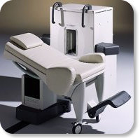
Developed by GE Lunar; the ARTOSCAN™-M is designed specifically for in-office musculoskeletal imaging. ARTOSCAN-M's compact, modular design allows placing within a clinical environment, bringing MRI to the patient. Patients remain outside the magnet at all times during the examinations, enabling constant patient-technologist contact. ARTOSCAN-M requires no special RF room, magnetic shielding, special power supply or air conditioning.
The C-SCAN™ (also known as Artoscan C) is developed from the ARTOSCAN™ - M, with a new computer platform.
Device Information and Specification
CLINICAL APPLICATION
Dedicated extremity
SE, GE, IR, STIR, FSE, 3D CE, GE-STIR, 3D GE, ME, TME, HSE
SLICE THICKNESS
2D: 2 mm - 10 mm;
3D: 0.6 mm - 10 mm
4,096 gray lvls, 256 lvls in 3D
POWER REQUIREMENTS
100/110/200/220/230/240V
| |  | |
• View the DATABASE results for 'ARTOSCAN™ - M' (3).
| | | | |
|  |  | Searchterm 'SPECIAL' was also found in the following services: | | | | |
|  |  |
| |
|
A pacemaker is a device for internal or external battery-operated cardiac pacing to overcome cardiac arrhythmias or heart block. All implanted electronic devices are susceptible to the electromagnetic fields used in magnetic resonance imaging. Therefore, the main magnetic field, the gradient field, and the radio frequency (RF) field are potential hazards for cardiac pacemaker patients.
The pacemaker's susceptibility to static field and its critical role in life support have warranted special consideration. The static magnetic field applies force to magnetic materials. This force and torque effects rise linearly with the field strength of the MRI machines. Both, RF fields and pulsed gradients can induce voltages in circuits or on the pacing lead, which will heat up the tissue around e.g. the lead tip, with a potential risk of thermal injury.
Regulations for pacemakers provide that they have to switch to the magnet mode in static magnetic fields above 1.0 mT. In MR imaging, the gradient and RF fields may mimic signals from the heart with inhibition or fast pacing of the heart. In the magnet mode, most of the current pacemakers will pace with a fix pulse rate because they do not accept the heartsignals. However, the state of an implanted pacemaker will be unpredictable inside a strong magnetic field. Transcutaneous controller adjustment of pacing rate is a feature of many units. Some achieve this control using switches activated by the external application of a magnet to open/close the switch. Others use rotation of an external magnet to turn internal controls. The fringe field around the MRI magnet can activate such switches or controls. Such activations are a safety risk.
Areas with fields higher than 0.5 mT ( 5 Gauss Limit) commonly have restricted access and/or are posted as a safety risk to persons with pacemakers.

A Cardiac pacemaker is because the risks, under normal circumstances an absolute contraindication for MRI procedures.
Nevertheless, with special precaution the risks can be lowered. Reprogramming the pacemaker to an asynchronous mode with fix pacing rate or turning off will reduce the risk of fast pacing or inhibition. Reducing the SAR value reduces the potential MRI risks of heating. For MRI scans of the head and the lower extremities, tissue heating also seems to be a smaller problem. If a transmit receive coil is used to scan the head or the feet, the cardiac pacemaker is outside the sending coil and possible heating is very limited. | |  | |
• View the DATABASE results for 'Cardiac Pacemaker' (6).
| | | | |  Further Reading: Further Reading: | Basics:
|
|
News & More:
| |
| |
|  | |  |  |  |
| |
|
Contrast agents are chemical substances introduced to the anatomical or functional region being imaged, to increase the differences between different tissues or between normal and abnormal tissue, by altering the relaxation times. MRI contrast agents are classified by the different changes in relaxation times after their injection.
•
Negative contrast agents (appearing predominantly dark on MRI) are small particulate aggregates often termed superparamagnetic iron oxide ( SPIO). These agents produce predominantly spin spin relaxation effects (local field inhomogeneities), which results in shorter T1 and T2 relaxation times.
SPIO's and ultrasmall superparamagnetic iron oxides ( USPIO) usually consist of a crystalline iron oxide core containing thousands of iron atoms and a shell of polymer, dextran, polyethyleneglycol, and produce very high T2 relaxivities. USPIOs smaller than 300 nm cause a substantial T1 relaxation. T2 weighted effects are predominant.
•
A special group of negative contrast agents (appearing dark on MRI) are perfluorocarbons ( perfluorochemicals), because their presence excludes the hydrogen atoms responsible for the signal in MR imaging.
The design objectives for the next generation of MR contrast agents will likely focus on prolonging intravascular retention, improving tissue targeting, and accessing new contrast mechanisms. Macromolecular paramagnetic contrast agents are being tested worldwide. Preclinical data shows that these agents demonstrate great promise for improving the quality of MR angiography, and in quantificating capillary permeability and myocardial perfusion.
Ultrasmall superparamagnetic iron oxide ( USPIO) particles have been evaluated in multicenter clinical trials for lymph node MR imaging and MR angiography, with the clinical impact under discussion. In addition, a wide variety of vector and carrier molecules, including antibodies, peptides, proteins, polysaccharides, liposomes, and cells have been developed to deliver magnetic labels to specific sites. Technical advances in MR imaging will further increase the efficacy and necessity of tissue-specific MRI contrast agents.
See also Adverse Reaction and Nephrogenic Systemic Fibrosis.
See also the related poll result: ' The development of contrast agents in MRI is' | | | | | | | | | | |
• View the DATABASE results for 'Contrast Agents' (122).
| | |
• View the NEWS results for 'Contrast Agents' (25).
| | | | |  Further Reading: Further Reading: | Basics:
|
|
News & More:
|  |
Brain imaging method may aid mild traumatic brain injury diagnosis
Tuesday, 16 January 2024 by parkinsonsnewstoday.com |  |  |
A Targeted Multi-Crystalline Manganese Oxide as a Tumor-Selective Nano-Sized MRI Contrast Agent for Early and Accurate Diagnosis of Tumors
Thursday, 18 January 2024 by www.dovepress.com |  |  |
FDA Approves Gadopiclenol for Contrast-Enhanced Magnetic Resonance Imaging
Tuesday, 27 September 2022 by www.pharmacytimes.com |  |  |
How to stop using gadolinium chelates for magnetic resonance imaging: clinical-translational experiences with ferumoxytol
Saturday, 5 February 2022 by www.ncbi.nlm.nih.gov |  |  |
Estimation of Contrast Agent Concentration in DCE-MRI Using 2 Flip Angles
Tuesday, 11 January 2022 by pubmed.ncbi.nlm.nih.gov |  |  |
Manganese enhanced MRI provides more accurate details of heart function after a heart attack
Tuesday, 11 May 2021 by www.news-medical.net |  |  |
Gadopiclenol: positive results for Phase III clinical trials
Monday, 29 March 2021 by www.pharmiweb.co |  |  |
Gadolinium-Based Contrast Agents Hypersensitivity: A Case Series
Friday, 4 December 2020 by www.dovepress.com |  |  |
Polysaccharide-Core Contrast Agent as Gadolinium Alternative for Vascular MR
Monday, 8 March 2021 by www.diagnosticimaging.com |  |  |
Water-based non-toxic MRI contrast agents
Monday, 11 May 2020 by chemistrycommunity.nature.com |  |  |
New method to detect early-stage cancer identified by Georgia State, Emory research team
Friday, 7 February 2020 by www.eurekalert.org |  |  |
Researchers Brighten Path for Creating New Type of MRI Contrast Agent
Friday, 7 February 2020 by www.newswise.com |  |  |
Manganese-based MRI contrast agent may be safer alternative to gadolinium-based agents
Wednesday, 15 November 2017 by www.eurekalert.org |  |  |
Sodium MRI May Show Biomarker for Migraine
Friday, 1 December 2017 by psychcentral.com |  |  |
A natural boost for MRI scans
Monday, 21 October 2013 by www.eurekalert.org |  |  |
For MRI, time is of the essence A new generation of contrast agents could make for faster and more accurate imaging
Tuesday, 28 June 2011 by scienceline.org |
|
| |
|  | |  |  |
|  | | |
|
| |
 | Look
Ups |
| |