 | Info
Sheets |
| | | | | | | | | | | | | | | | | | | | | | | | |
 | Out-
side |
| | | | |
|
| | | | |
Result : Searchterm 'Rephasing' found in 6 terms [ ] and 14 definitions [ ] and 14 definitions [ ] ]
| | previous 11 - 15 (of 20) nextResult Pages : [1 2] [3 4] |  | |  | Searchterm 'Rephasing' was also found in the following service: | | | | |
| |  |
| |
|
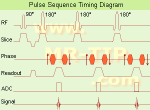
(FSE) In the pulse sequence timing diagram, a fast spin echo sequence with an echo train length of 3 is illustrated.
This sequence is characterized by a series of rapidly applied 180° rephasing pulses and multiple echoes, changing the phase encoding gradient for each echo.
The echo time TE may vary from echo to echo in the echo train. The echoes in the center of the K-space (in the case of linear k-space acquisition) mainly produce the type of image contrast, whereas the periphery of K-space determines the spatial resolution. For example, in the middle of K-space the late echoes of T2 weighted images are encoded. T1 or PD contrast is produced from the early echoes.
The benefit of this technique is that the scan duration with, e.g. a turbo spin echo turbo factor / echo train length of 9, is one ninth of the time. In T1 weighted and proton density weighted sequences, there is a limit to how large the ETL can be (e.g. a usual ETL for T1 weighted images is between 3 and 7). The use of large echo train lengths with short TE results in blurring and loss of contrast. For this reason, T2 weighted imaging profits most from this technique.
In T2 weighted FSE images, both water and fat are hyperintense. This is because the succession of 180° RF pulses reduces the spin spin interactions in fat and increases its T2 decay time. Fast spin echo (FSE) sequences have replaced conventional T2 weighted spin echo sequences for most clinical applications. Fast spin echo allows reduced acquisition times and enables T2 weighted breath hold imaging, e.g. for applications in the upper abdomen.
In case of the acquisition of 2 echoes this type of a sequence is named double fast spin echo / dual echo sequence, the first echo is usually density and the second echo is T2 weighted image. Fast spin echo images are more T2 weighted, which makes it difficult to obtain true proton density weighted images. For dual echo imaging with density weighting, the TR should be kept between 2000 - 2400 msec with a short ETL (e.g., 4).
Other terms for this technique are:
Turbo Spin Echo
Rapid Imaging Spin Echo,
Rapid Spin Echo,
Rapid Acquisition Spin Echo,
Rapid Acquisition with Refocused Echoes
| | | | | | | | | | | | |  Further Reading: Further Reading: | | Basics:
|
|
News & More:
| |
| |
| | | | | |
| |
|
Quick Overview Please note that there are different common names for this artifact.
REASON
Movement of body fluids
Flow effects in MRI produce a range of artifacts, e.g. intravascular signal void by time of flight effects; turbulent dephasing and first echo dephasing, caused by flowing blood.
Through movement of the hydrogen nuclei (e.g. blood flow), there is a location change between the time these nuclei experience a radio frequency pulse and the time the emitted signal is received (because the repetition time is asynchronous with the pulsatile flow).
The blood flow occasionally produces intravascular high signal intensities due to flow related enhancement, even echo rephasing and diastolic pseudogating. The pulsatile laminar flow within vessels often produces a complex multilayered band that usually propagates outside the head in the phase encoded direction. Blood flow artifacts should be considered as a special subgroup of motion artifacts.

Image Guidance
| | | | | |
• View the DATABASE results for 'Flow Artifact' (6).
| | | | | | Further Reading: | News & More:
|
|
| |
| | | | | |
| |
|
Quick Overview
NAME
Metal, susceptibility
Ferromagnetic metal will cause a magnetic field inhomogeneity, which in turn causes a local signal void, often accompanied by an area of high signal intensity, as well as a distortion of the image.
They create their own magnetic field and dramatically alter precession frequencies of protons in the adjacent tissues. Tissues adjacent to ferromagnetic components become influenced by the induced magnetic field of the metal hardware rather than the parent field and, therefore, either fail to precess or do so at a different frequency and hence do not generate useful signal. Two components contribute to susceptibility artifact, induced magnetism in the ferromagnetic component itself and induced magnetism in protons adjacent to the component. Artifacts from metal may have varied appearances on MRI scans due to different type of metal or configuration of the piece of metal.
The biocompatibility of metallic alloys, stainless steel, cobalt chrome and titanium alloy is based on the presence of a constituent element within the alloy that has the ability to form an adherent oxide coating that is stable, chemically inert and hence biocompatible. In relation to imaging titanium alloys are less ferromagnetic than both cobalt and stainless steel, induce less susceptibility artifact and result in less marked image degradation.
Image Guidance
| | | |
• View the DATABASE results for 'Metal Artifact' (2).
| | | | | | Further Reading: | | Basics:
|
|
News & More:
| |
| |
| | | Searchterm 'Rephasing' was also found in the following service: | | | | |
| | |
| |
|
Quick Overview
Please note that there are different common names for this artifact.
NAME
Motion, phase encoded motion, instability, smearing
REASON
Movement of the imaged object
HELP
Compensation techniques, more averages, anti spasmodic
Patient motion is the largest physiological effect that causes artifacts, often resulting from involuntary movements (e.g. respiration, cardiac motion and blood flow, eye movements and swallowing) and minor subject movements.
Movement of the object being imaged during the sequence results in inconsistencies in phase and amplitude, which lead to blurring and ghosting. The nature of the artifact depends on the timing of the motion with respect to the acquisition. Causes of motion artifacts can also be mechanical vibrations, cryogen boiling, large iron objects moving in the fringe field (e.g. an elevator), loose connections anywhere, pulse timing variations, as well as sample motion. These artifacts appear in the phase encoding direction, independent of the direction of the motion.
Image Guidance
| | | |
• View the DATABASE results for 'Motion Artifact' (24).
| | | | | | Further Reading: | Basics:
|
|
News & More:
| |
| |
| | | | | |
| |
|
| | | |
• View the DATABASE results for 'Motion Artifact Suppression Technique' (3).
| | | | | | Further Reading: | News & More:
|
|
| |
| | | | |
| | | |
|
| |
 | Look
Ups |
| |


