Magnetic Resonance - Technology Information Portal Welcome to MRI Technology
Info
  Sheets

Out-
      side


 'Diffusion Time' 
SEARCH FOR    
 
  2 3 5 A B C D E F G H I J K L M N O P Q R S T U V W X Y Z
Result : Searchterm 'Diffusion Time' found in 1 term [] and 1 definition [], (+ 17 Boolean[] results
previous     6 - 10 (of 19)     next
Result Pages : [1]  [2 3 4]
Searchterm 'Diffusion Time' was also found in the following services: 
spacer
News  (2)  Resources  (1)  
 
Ultra™InfoSheet: - Devices -
Intro, 
Types of Magnets, 
Overview, 
etc.MRI Resource Directory:
 - Devices -
 
medical.toshiba.com/clinical/radiology/ultra.htm 
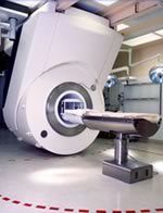
From Toshiba America Medical Systems Inc.;
the Ultra™ system was developed to help healthcare providers be more competitive by delivering greater patient comfort and a broad range of clinical capabilities, says Anita Bowler, product manager, MRI Business Unit, Toshiba America Medical Systems Inc With its unique, powerful gradient technology, the Ultra™ performs advanced clinical studies and consistently provides high-resolution images that are typically associated with high field MRI systems. At the same time, the Ultra™ offers a truly open feeling that makes patients more relaxed, especially those with claustrophobic tendencies.
Device Information and Specification
CLINICAL APPLICATION
Whole Body
CONFIGURATION
Quadrature, solenoid and multi-channel configurations
PULSE SEQUENCES
SE, FE, IR, FastSE, FastIR, FastFLAIR, Fast STIR, FastFE, FASE, Hybrid EPI, Multi Shot EPI, Single shot EPI diffusion, True SSFP, SuperFASE; Angiography: 2D(gate/non-gate)/3D TOF, SORS-STC, Black Blood MRA
POWER REQUIREMENTS
380/400/415/440/480 V
COOLING SYSTEM TYPE
Cryogenless
STRENGTH
25 mT/m
Passive, active
spacer
MRI Resources 
Image Quality - Pediatric and Fetal MRI - Contrast Enhanced MRI - Examinations - Pregnancy - Spectroscopy
 
iMotion™ 1.5 Tesla MagnetInfoSheet: - Devices -
Intro, 
Types of Magnets, 
Overview, 
etc.MRI Resource Directory:
 - Devices -
 
www.imris.com/ The mobile intraoperative iMotion system produces real-time images used for MR guided surgery and offers functional magnetic resonance imaging, MR spectroscopy, perfusion imaging, and diffusion weighted imaging capabilities.
The iMotion 1.5 T magnet moves to the patient, gliding in and out of place as needed, without affecting surgical, anesthetic, and nursing management.

See also Intraoperative Magnetic Resonance Imaging, MR Guided Interventions.
Device Information and Specification
CLINICAL APPLICATION
Whole body
CONFIGURATION
Mobile compact
Whole body, intra-operative head, neck volume, atlas head//neck vascular quadrature phased array, spine quadrature, C/T/L spine phased array, small joint, large joint, TMJ bilateral, shoulder phased array, extremity quadrature volume, wrist, hand quadrature, general purpose flexible, pelvis/abdomen phased array, body quadrature, phased array flexible, breast bilateral
SYNCHRONIZATION
Standard cardiac gating, ECG/peripheral, respiratory gating
PULSE SEQUENCES
SE, IR, TSE, EPI, Diffusion, Perfusion, Angiography
IMAGING MODES
Localizer, single slice, multislice, volume
Active
spacer

• View the DATABASE results for 'iMotion™ 1.5 Tesla Magnet' (2).Open this link in a new window

MRI Resources 
DICOM - Resources - Stimulator pool - Anatomy - RIS - Education pool
 
CHORUS 1.5T™InfoSheet: - Devices -
Intro, 
Types of Magnets, 
Overview, 
etc.MRI Resource Directory:
 - Devices -
 
www.isoltech.co.kr/english/product/default.htm 
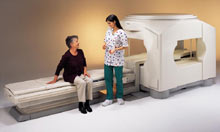
'Next generation MRI system 1.5T CHORUS developed by ISOL Technology is optimized for both clinical diagnostic imaging and for research development.
CHORUS offers the complete range of feature oriented advanced imaging techniques- for both clinical routine and research. The compact short bore magnet, the patient friendly design and the gradient technology make the innovation to new degree of perfection in magnetic resonance.'
Device Information and Specification
CLINICAL APPLICATION
Whole body
CONFIGURATION
Short bore
Head, C-spine, L-spine, TMJ, Knee, Shoulder, General purpose, Phased Array System: 4 digital receiver channels (Up to 12 channels)
Optional
SYNCHRONIZATION
ECG/peripheral: Optional/yes, respiratory gating
PULSE SEQUENCES
Spin Echo, Gradient Echo, Fast Spin Echo, Inversion Recovery (STIR, Fluid Attenuated Inversion Recovery), FLASH, FISP, PSIF, Turbo Flash ( MPRAGE ),TOF MR Angiography, Standard echo planar imaging package (SE-EPI, GE-EPI), Optional: Advanced P.A. Imaging Package (up to 4 ch.), Advanced echo planar imaging package, Single Shot and Diffusion Weighted EPI, IR/FLAIR EPI
IMAGING MODES
2D/3D, Travelling Sat, Multi-Slab 3D, MTC and TONE Pulse Sequence, Fat/Water Suppression, Presaturation (up to 6 bands), Flow Compensation using GMR pulse, Multi-Slice, Multi-Group Imaging
SINGLE/MULTI SLICE
Image reconstruction time (2562 ) : 0.02 s
FOV
40 cm
BORE DIAMETER
or W x H
58 cm diameter (patient)
MAGNET WEIGHT
4050 kg
H*W*D
233 x 206 x 160 cm
COOLING SYSTEM TYPE
Water-cooled coil and air-cooled amplifier
CRYOGEN USE
0.07 L/hr helium
STRENGTH
20 mT/m (Upto 27 mT/m)
5-GAUSS FRINGE FIELD
2.5 m / 3.8 m
Passive and active
spacer

• View the DATABASE results for 'CHORUS 1.5T™' (2).Open this link in a new window

Searchterm 'Diffusion Time' was also found in the following services: 
spacer
News  (2)  Resources  (1)  
 
Lung ImagingMRI Resource Directory:
 - Lung Imaging -
 
Lung imaging is furthermore a challenge in MRI because of the predominance of air within the lungs and associated susceptibility issues as well as low signal to noise of the inflated lung parenchyma. Cardiac and respiratory triggered or breath hold sequences allow diagnostic imaging, however a comparable image quality with computed tomography is still difficult to achieve.
Assumptions for lung MRI:
•
Low signal to noise ratio of the inherently low lung proton density.
•
Cardiac and respiratory motion artifacts.
•
Magnetic susceptibility effects of large magnetic field gradients.
•
Very short transverse relaxation times and significant diffusion yielding short T2 (30-70 msec), short T2* (1-3 msec), and additional long T1 relaxation times (1300-1500 msec).
•
The extreme short T2 values are responsible for a fast signal decay during a single shot readout, resulting in blurring.

The current trends in MRI are the use of new imaging technologies and increasingly powerful magnetic fields. Among these technologies are parallel imaging techniques as well as ventilation agents like hyperpolarized helium for the use as an inert inhalational contrast agent to study lung ventilation properties. With hyperpolarized gases clear images of the lungs can be obtained without using a large magnetic field (see also back projection imaging). Single shot sequences (e.g. TSE or Half Fourier Acquisition Single Shot Turbo Spin Echo HASTE) used in lung MR imaging benefits from parallel imaging techniques due to reduced relaxation time effects during the echo train and therefore reduced image blurring as well as reduced motion artifacts.
In the future, more effective contrast agents may provide an alternative solution to the need for high field MRI. Dynamic contrast enhanced MRI perfusion has demonstrated a potential in the diagnosis of pulmonary embolism or to characterize lung cancer and mediastinal tumors. 3D contrast enhanced magnetic resonance angiography of the thoracic vessel.

See also the related poll result: 'MRI will have replaced 50% of x-ray exams by'
 
Images, Movies, Sliders:
 Anatomic Imaging of the Lungs  Open this link in a new window
      

Courtesy of  Robert R. Edelman
 Normal Lung Gd Perfusion MRI  Open this link in a new window
      

Courtesy of  Robert R. Edelman

 MRI Thorax Basal Plane  Open this link in a new window
 
Radiology-tip.comradLung Scintigraphy
spacer

• View the DATABASE results for 'Lung Imaging' (7).Open this link in a new window


• View the NEWS results for 'Lung Imaging' (3).Open this link in a new window.
 
Further Reading:
  Basics:
A safer approach for diagnostic medical imaging
Monday, 29 September 2014   by www.eurekalert.org    
Parallel Lung Imaging(.pdf)
  News & More:
Chest MRI a viable alternative to chest CT in COVID-19 pneumonia follow-up
Monday, 21 September 2020   by www.healthimaging.com    
CT Imaging Features of 2019 Novel Corona virus (2019-nCoV)
Tuesday, 4 February 2020   by pubs.rsna.org    
Polarean Imaging Phase III Trial Results Point to Potential Improvements in Lung Imaging
Wednesday, 29 January 2020   by www.diagnosticimaging.com    
Low Power MRI Helps Image Lungs, Brings Costs Down
Thursday, 10 October 2019   by www.medgadget.com    
Chest MRI Using Multivane-XD, a Novel T2-Weighted Free Breathing MR Sequence
Thursday, 11 July 2019   by www.sciencedirect.co    
Researchers Review Importance of Non-Invasive Imaging in Diagnosis and Management of PAH
Wednesday, 11 March 2015   by lungdiseasenews.com    
New MRI Approach Reveals Bronchiectasis' Key Features Within the Lung
Thursday, 13 November 2014   by lungdiseasenews.com    
MRI techniques improve pulmonary embolism detection
Monday, 19 March 2012   by medicalxpress.com    
  News & More:
Partnership with VIDA to streamline adoption of advanced MRI of the lungs
Monday, 11 September 2023   by www.itnonline.com    
MRI Resources 
Calculation - Bioinformatics - Breast MRI - Musculoskeletal and Joint MRI - Spine MRI - Pregnancy
 
Intravoxel Incoherent Motion
 
(IVIM) Spins moving in fluids with different velocities and possibly in different directions. This is being found to a small degree in all tissues as a result of capillary perfusion or diffusion. Important velocity changes occur as one moves from the vessel wall towards the center of the vessel. Hence, spins (to a variable degree) have different velocities within a single imaging voxel.
This effect can be measured using special pulse sequences such as in diffusion imaging or diffusion weighed imaging. When the velocity differences are marked, as occurs in larger blood vessels, effects due to IVIM are visible in standard MR images and give rise to flow related dephasing. The effects are more visible when longer echo times are used.
spacer

• View the DATABASE results for 'Intravoxel Incoherent Motion' (3).Open this link in a new window

 
Further Reading:
  Basics:
Diffusion Imaging: From Basic Physics to Practical Imaging
1999   by ej.rsna.org    
  News & More:
EVALUATION OF HUMAN STROKE BY MR IMAGING
2000
MRI Resources 
IR - Shielding - MRI Reimbursement - Quality Advice - Abdominal Imaging - Veterinary MRI
 
previous      6 - 10 (of 19)     next
Result Pages : [1]  [2 3 4]
 Random Page
 
Share This Page
FacebookTwitterLinkedIn

MR-TIP    
Community   
User
Pass
Forgot your UserID/Password ?    


New acceleration techniques will :
reduce scan times 
cause artifacts 
increase expenses 
be useful if you have a lot of experience 
doesn't do much 
never heard of 

Look
      Ups


MR-TIP.com uses cookies! By browsing MR-TIP.com, you agree to our use of cookies.

Magnetic Resonance - Technology Information Portal
Member of SoftWays' Medical Imaging Group - MR-TIP • Radiology-TIP • Medical-Ultrasound-Imaging • 
Copyright © 2003 - 2024 SoftWays. All rights reserved. [ 26 April 2024]
Terms of Use | Privacy Policy | Advertising
 [last update: 2024-02-26 03:41:00]