 | Info
Sheets |
| | | | | | | | | | | | | | | | | | | | | | | | |
 | Out-
side |
| | | | |
|
| | | | |
Result : Searchterm 't1 stir' found in 0 term [ ] and 0 definition [ ] and 0 definition [ ], (+ 20 Boolean[ ], (+ 20 Boolean[ ] results ] results
| | previous 11 - 15 (of 20) nextResult Pages : [1 2 3 4] |  | |  | Searchterm 't1 stir' was also found in the following service: | | | | |
| |  |
| |
|
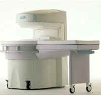
Device Information and Specification
CLINICAL APPLICATION
Whole body
CONFIGURATION
Short bore compact
Standard: Head, body, cardiac, optional phased array: Spine, pediatric, 3rd party connector; Optional SENSE? coils for all applications
SE, Modified-SE, IR ( T1, T2, PD), STIR, FLAIR, SPIR, FFE, T1-FFE, T2-FFE, Balanced FFE, TFE, Balanced TFE, Dynamic, Keyhole, 3D, Multi Chunk 3D, Multi Stack 3D, K Space Shutter, MTC, TSE, Dual IR, DRIVE, EPI, Cine, 2DMSS, DAVE, Mixed Mode; Angiography: Inflow MRA, TONE, PCA, CE MRA
128 x 128, 256 x 256,512 x 512,1024 x 1024 (64 for Bold img)
Variable in 1% increments
Lum.: 120 cd/m2; contrast: 150:1
Variable (op. param. depend.)
POWER REQUIREMENTS
380/400 V
| | | | | |  Further Reading: Further Reading: | News & More:
|
|
| |
| | | | | |
| |
|
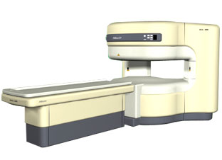
Device Information and Specification
CLINICAL APPLICATION
Whole body
CONFIGURATION
Short bore compact
Standard: head, body, C1, C3; Optional: Small joint, flex-E, flex-R, endocavitary (L and S), dual TMJ, knee, neck, T/L spine, breast; Optional phased array: Spine, pediatric, 3rd party connector, Optional SENSE Coils: Flex-S-M-L, Flex Body, Flex Cardiac
SE, Modified-SE, IR ( T1, T2, PD), STIR, FLAIR, SPIR, FFE, T1-FFE, T2-FFE, Balanced FFE, TFE, Balanced TFE, Dynamic, Keyhole, 3D, Multi Chunk 3D, Multi Stack 3D, K Space Shutter, MTC, TSE, Dual IR, DRIVE, EPI, Cine, 2DMSS, DAVE, Mixed Mode; Angiography: Inflow MRA, TONE, PCA, CE MRA
TR
Min. 2.9 (Omni) msec, 1.6 (Power) msec
TE
Min. 1.0 (Omni) msec, 0.7 (Power) msec
RapidView Recon. greater than 500 @ 256 Matrix
0.1 mm(Omni), 0.05 mm (Power)
128 x 128, 256 x 256,512 x 512,1024 x 1024 (64 for Bold img)
Variable in 1% increments
Lum.: 120 cd/m2; contrast: 150:1
Variable (op. param. depend.)
POWER REQUIREMENTS
380/400 V
STRENGTH
23 mT/m (Omni), 30 (Power) mT/m
| | | |
• View the DATABASE results for 'Intera 1.0T™' (2).
| | | | |
| | | | | |
| |
|
From Siemens Medical Systems;
This open MRI system is a dedicated extremity scanner, which is easy to use and easy to install. Whether you want to use it as a supplement to your whole-body scanner or you want to specialize in orthopedic exams,
MAGNETOM Jazz™ offers you a cost effective opportunity to make the most of your capabilities.
Device Information and Specification GRE, IR, FIR, STIR, TrueIR/FISP, FSE, MT, SS-FSE, MT-SE, MTC, MSE, GMR, fat/water sat./exc. IMAGING MODES Single, multislice, volume study, multi angle 512 x 512 full screen display POWER REQUIREMENTS 380/400/420/440/480 V | | | |
• View the DATABASE results for 'MAGNETOM Jazz™' (2).
| | | | |
| | | Searchterm 't1 stir' was also found in the following service: | | | | |
| | |
| |
|
From ISOL Technology
'RELAX is open type MRI system created by making up for the weakness of existing conventional MR systems and applying the strength and the application of the middle to high field MR without uncompromising the image quality.
RELAX offers you a premium mix of form, performance and functionality that are patient and user
friendly beyond comparison.
- New breed of MRI pursuing
- patients comfort'
Device Information and Specification CLINICAL APPLICATION Whole body lower than 2.4 m from the iso-center | | | |
• View the DATABASE results for 'RELAX 0.35T™' (2).
| | | | |
| | | | | |
| |
|
( STIR) Also called Short Tau ( t) ( inversion time) Inversion Recovery. STIR is a fat suppression technique with an inversion time t = T1 ln2 where the signal of fat is zero ( T1 is the spin lattice relaxation time of the component that should be suppressed). To distinguish two tissue components with this technique, the T1 values must be different. Fluid Attenuation Inversion Recovery ( FLAIR) is a similar technique to suppress water.
Inversion recovery doubles the distance spins will recover, allowing more time for T1 differences. A 180° preparation pulse inverts the net magnetization to the negative longitudinal magnetization prior to the 90° excitation pulse.
This specialized application of the inversion recovery sequence set the inversion time ( t) of the sequence at 0.69 times the T1 of fat. The T1 of fat at 1.5 Tesla is approximately 250 with a null point of 170 ms while at 0.5 Tesla its 215 with a 148 ms null point. At the moment of excitation, about 120 to 170 ms after the 180° inversion pulse (depending of the magnetic field) the magnetization of the fat signal has just risen to zero from its original, negative, value and no fat signal is available to be flipped into the transverse plane.
When deciding on the optimal T1 time, factors to be considered include not only the main field strength, but also the tissue to be suppressed and the anatomy. In comparison to a conventional spin echo where tissues with a short T1 are bright due to faster recovery, fat signal is reversed or darkened.
Because body fluids have both a long T1 and a long T2, it is evident that STIR offers the possibility of extremely sensitive detection of body fluid. This is of course, only true for stationary fluid such as edema, as the MRI signal of flowing fluids is governed by other factors.
See also Fat Suppression and Inversion Recovery Sequence. | | | | | |
• View the DATABASE results for 'Short T1 Inversion Recovery' (3).
| | | | | | Further Reading: | | Basics:
|
|
News & More:
| |
| |
| | | | |
| | |
| | | |
|
| |
 | Look
Ups |
| |


