| Info
Sheets |
| | | | | | | | | | | | | | | | | | | | | | | | |
 | Out-
side |
| | | | |
|
| | | | |
Result : Searchterm 'Array' found in 8 terms [ ] and 53 definitions [ ] and 53 definitions [ ] ]
| | previous 16 - 20 (of 61) nextResult Pages : [1 2] [3 4 5 6 7 8 9 10 11 12 13] |  | |  | Searchterm 'Array' was also found in the following services: | | | | |
| |  |
| |
|
(SENSE) A MRI technique for relevant scan time reduction. The spatial information related to the coils of a receiver array are utilized for reducing conventional Fourier encoding. In principle, SENSE can be applied to any imaging sequence and k-space trajectories. However, it is particularly feasible for Cartesian sampling schemes. In 2D Fourier imaging with common Cartesian sampling of k-space sensitivity encoding by means of a receiver array enables to reduce the number of Fourier encoding steps.
SENSE reconstruction without artifacts relies on accurate knowledge of the individual coil sensitivities. For sensitivity assessment, low-resolution, fully Fourier-encoded reference images are required, obtained with each array element and with a body coil.
The major negative point of parallel imaging techniques is that they diminish SNR in proportion to the numbers of reduction factors.
R is the factor by which the number of k-space samples is reduced. In standard Fourier imaging reducing the sampling density results in the reduction of the FOV, causing aliasing. In fact, SENSE reconstruction in the Cartesian case is efficiently performed by first creating one such aliased image for each array element using discrete Fourier transformation (DFT).
The next step then is to create a full-FOV image from the set of intermediate images. To achieve this one must undo the signal superposition underlying the fold-over effect. That is, for each pixel in the reduced FOV the signal contributions from a number of positions in the full FOV need to be separated. These positions form a Cartesian grid corresponding to the size of the reduced FOV.
The advantages are especially true for contrast-enhanced MR imaging such as
dynamic liver MRI (liver imaging) ,
3 dimensional magnetic resonance angiography (3D MRA), and magnetic resonance cholangiopancreaticography ( MRCP).
The excellent scan speed of SENSE allows for acquisition of two separate sets of hepatic MR images within the time regarded as the hepatic arterial-phase (double arterial-phase technique) as well as that of multidetector CT.
SENSE can also increase the time efficiency of spatial signal encoding in 3D MRA. With SENSE, even ultrafast (sub second) 4D MRA can be realized.
For MRCP acquisition, high-resolution 3D MRCP images can be constantly provided by SENSE. This is because SENSE resolves the presence of the severe motion artifacts due to longer acquisition time. Longer acquisition time, which results in diminishing image quality, is the greatest problem for 3D MRCP imaging.
In addition, SENSE reduces the train of gradient echoes in combination with a faster k-space traversal per unit time, thereby dramatically improving the image quality of single shot echo planar imaging (i.e. T2 weighted, diffusion weighted imaging). | | | | | | | | | | |  Further Reading: Further Reading: | News & More:
|
|
| |
| | | | | |
| |
|
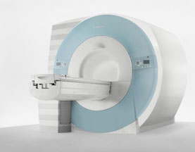
Device Information and Specification
CLINICAL APPLICATION
Whole body
CONFIGURATION
Cylindrical Wide Short Bore
Opt. (WIP) Single and Multi Voxel
SE, FE, IR, FastSE, FastIR, FastFLAIR, Fast STIR, FastFE, FASE, Hybrid EPI, Multi Shot EPI; Angiography: 2D(gate/non-gate)/3D TOF, SORS-STC
IMAGING MODES
Single, multislice, volume study
TE
8 msec min. SE; 1.2 msec min. FE
less than 0.015 (256x256)
1.0 min. 2-DFT: 0.2 min. 3-DFT
32-1024, phase;; 64-1024, freq.
65.5 cm, patient aperture
4050 kg (bare magnet incl. L-He)
COOLING SYSTEM TYPE
Closed-loop water-cooled
Liquid helium: approx. less than 0.05 L/hr
Passive, active, auto-active
| | | |
• View the DATABASE results for 'Excelart AG™ with Pianissimo' (2).
| | | | |
| | | | | |
| |
|
Device Information and Specification
CLINICAL APPLICATION
Whole body
CONFIGURATION
Cylindrical Wide Short Bore
SE, FE, IR, FastSE, FastIR, FastFLAIR, Fast STIR, FastFE, FASE, Hybrid EPI, Multi Shot EPI; Angiography: 2D(gate/non-gate)/3D TOF, SORS-STC
IMAGING MODES
Single, multislice, volume study
TE
8 msec min. SE; 0.9 msec min. FE
less than 0.011 (256x256)
1.0 min. 2-DFT: 0.2 min. 3-DFT
32-1024, phase;; 64-1024, freq.
65.5 cm, patient aperture
4050 kg (bare magnet incl. L-He)
POWER REQUIREMENTS
380/400/415/440/480 V
COOLING SYSTEM TYPE
Closed-loop water-cooled
Liquid helium: approx. less than 0.05 L/hr
Passive, active, auto-active
| | | |
• View the DATABASE results for 'Excelart XG™ with Pianissimo' (2).
| | | | | | Further Reading: | News & More:
|
|
| |
| | | Searchterm 'Array' was also found in the following services: | | | | |
| | |
| |
|

From Esaote S.p.A.;
Esaote introduced the new G-SCAN at the RSNA in Dec. 2004. The G-SCAN covers almost all musculoskeletal applications including the spine. The tilting gantry is designed for scanning in weight-bearing positions. This unique MRI scanner is developed in line with the Esaote philosophy of creating high quality MRI systems that are easy to install and that have a low breakeven point.
Device Information and Specification
SE, GE, IR, STIR, TSE, 3D CE, GE-STIR, 3D GE, ME, TME, HSE
100 up to 350 mm, 25 mm displayed
POWER REQUIREMENTS
100/110/200/220/230/240 V
| | | |
• View the DATABASE results for 'G-SCAN' (3).
| | | | |
| | | | | |
| |
|
Device Information and Specification CLINICAL APPLICATION Whole body SE, IR, 2D/3D TurboSE, Turbo IR, Dark-Fluid IR, True IR, 2D/3D MEDIC, 2D/3D GRE FLASH, 2D/3D GRE FISP, 2D/3D PSIF, 2D TurboFLASH, 3D MP-RAGE, 3D TurboFLASH, 2D/3D TOF angiography, MTC, TONE with 3D TOF MRA, GMR, LOTA IMAGING MODES Single, multislice, volume study, multi angle, multi oblique178 images/sec at 256 x 256 at 100% FOV1024 x 1024 full screen display POWER REQUIREMENTS 380/400/420/440/480 V Passive, act.; 1st order std./2nd opt. | | | | | |
| | | | |
| | |
| | | |
|
| |
 | Look
Ups |
| |