 | Info
Sheets |
| | | | | | | | | | | | | | | | | | | | | | | | |
 | Out-
side |
| | | | |
|
| | | | |
Result : Searchterm 'T1 Weighted Image' found in 1 term [ ] and 17 definitions [ ] and 17 definitions [ ], (+ 14 Boolean[ ], (+ 14 Boolean[ ] results ] results
| previous 16 - 20 (of 32) nextResult Pages :  [1] [1]  [2 3 4] [2 3 4]  [5 6 7] [5 6 7] |  | |  | Searchterm 'T1 Weighted Image' was also found in the following services: | | | | |
|  |  |
| |
|
(PS) Excitation technique applying repeated RF pulses in times comparable to or shorter than T1.
Incomplete T1 relaxation leads to reduction of the signal amplitude; there is the possibility of generating images with increased contrast between regions with different relaxation times.
Although partial saturation is also commonly referred to as saturation recovery, that term should properly be reserved for the particular case of partial saturation in which recovery after each excitation effectively takes place from true saturation.
A GRE sequence where ╬▒ = 90┬░ is identical to the partial saturation or saturation recovery pulse sequence.
It does not directly produce images of T1. However, since the measured signal will depend on T1, the method generates contrast between regions with different relaxation times. If T2 and/or T2 effects are minimized through the use of a short echo time TE, the result is a T1 weighted image. It is not a T1 image due to the possible presence of spin density and T2 effects as well as the nonlinear dependence on T1.
The change in signal from a region resulting from a change in the interpulse time, TR, can be used to calculate T1 for the region. | |  | | | | | | | | |
|  | |  |  |  |
| |
|
Resovist® is an organ-specific MRI contrast agent, used for the detection and characterization of especially small focal liver lesions.
Resovist® consists of superparamagnetic iron oxide ( SPIO) nanoparticles coated with carboxydextran, which are accumulated by phagocytosis in cells of the reticuloendothelial system (RES) of the liver. The uptake of Resovist® Injection in the reticuloendothelial cells results in a decrease of the signal intensity of normal liver parenchyma on both T2- and T1 weighted images.
Most malignant liver tumors do not contain RES cells and therefore do not uptake the iron particles. The resulting imaging effect is an improved contrast between the tumor (bright) and the surrounding tissue (dark).
Resovist® can be injected as an intravenous bolus, which allows immediate imaging of the liver and reduces the overall examination time. A dynamic imaging strategy after bolus injection supports to characterize lesions.
In comprehensive clinical trials, it demonstrated an excellent safety profile.
In 2001, Resovist® was approved for the European market.
See also Superparamagnetic Iron Oxide.
Resovist┬« competed with PrimovistÔäó, the other liver imaging agent of Bayer Schering Pharma AG. Due to this reason, the production of Resovist┬« has been abandoned in 2009. Drug Information and Specification T2/T1, Predominantly negative enhancement PHARMACOKINETIC RES-directed CONCENTRATION 0.5 mol Fe/L DOSAGE Less than 60 kg = 0.9 ml, greater than 60 kg = 1.4 ml PREPARATION Finished product PRESENTATION
Pre-filled syringes of 0.9 and 1.4 mL DO NOT RELY ON THE INFORMATION PROVIDED HERE, THEY ARE
NOT A SUBSTITUTE FOR THE ACCOMPANYING PACKAGE INSERT! Distribution Information TERRITORY TRADE NAME DEVELOPMENT
STAGE DISTRIBUTOR Japan Resovist® approved - Australia Resovist® Approved - | |  | |
• View the DATABASE results for 'Resovist®' (6).
| | | | |  Further Reading: Further Reading: | News & More:
|
|
| |
|  | |  |  |  |
| |
|
Signal intensity interpretation in MR imaging has a major problem.
Often there is no intuitive approach to signal behavior as signal intensity is a very complicated function of the contrast-determining tissue parameter, proton density, T1 and T2, and the machine parameters TR and TE. For this reason, the terms T1 weighted image, T2 weighted image and proton density weighted image were introduced into clinical MR imaging.
Air and bone produce low-intensity, weaker signals with darker images. Fat and marrow produce high-intensity signals with brighter images.
The signal intensity measured is related to the square of the xy-magnetization, which in a SE pulse sequence is given by
Mxy = Mxy0(1-exp(-TR/T1)) exp(-TE/T2) (1)
where Mxy0 = Mz0 is proportional to the proton or spin density, and corresponds to the z-magnetization present at zero time of the experiment when it is tilted into the xy-plane. See also T2 Weighted Image and Ernst Angle. | |  | |
• View the DATABASE results for 'Signal Intensity' (56).
| | |
• View the NEWS results for 'Signal Intensity' (1).
| | | | |  Further Reading: Further Reading: | | Basics:
|
|
News & More:
| |
| |
|  |  | Searchterm 'T1 Weighted Image' was also found in the following services: | | | | |
|  |  |
| |
|

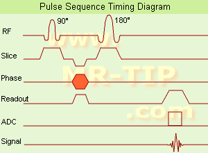
(SE) The most common pulse sequence used in MR imaging is based of the detection of a spin or Hahn echo. It uses 90┬░ radio frequency pulses to excite the magnetization and one or more 180┬░ pulses to refocus the spins to generate signal echoes named spin echoes (SE).
In the pulse sequence timing diagram, the simplest form of a spin echo sequence is illustrated.
The 90┬░ excitation pulse rotates the longitudinal magnetization ( Mz) into the xy-plane and the dephasing of the transverse magnetization (Mxy) starts.
The following application of a 180° refocusing pulse (rotates the magnetization in the x-plane) generates signal echoes. The purpose of the 180° pulse is to rephase the spins, causing them to regain coherence and thereby to recover transverse magnetization, producing a spin echo.
The recovery of the z-magnetization occurs with the T1 relaxation time and typically at a much slower rate than the T2-decay, because in general T1 is greater than T2 for living tissues and is in the range of 100-2000 ms.
The SE pulse sequence was devised in the early days of NMR days by Carr and Purcell and exists now in many forms: the multi echo pulse sequence using single or multislice acquisition, the fast spin echo (FSE/TSE) pulse sequence, echo planar imaging (EPI) pulse sequence and the gradient and spin echo (GRASE) pulse sequence;; all are basically spin echo sequences.
In the simplest form of SE imaging, the pulse sequence has to be repeated as many times as the image has lines. Contrast values:
PD weighted: Short TE (20 ms) and long TR.
T1 weighted: Short TE (10-20 ms) and short TR (300-600 ms)
T2 weighted: Long TE (greater than 60 ms) and long TR (greater than 1600 ms)
With spin echo imaging no T2* occurs, caused by the 180┬░ refocusing pulse. For this reason, spin echo sequences are more robust against e.g., susceptibility artifacts than gradient echo sequences.
See also Pulse Sequence Timing Diagram to find a description of the components.
| | | |  | |
• View the DATABASE results for 'Spin Echo Sequence' (24).
| | | | |  Further Reading: Further Reading: | Basics:
|
|
News & More:
| |
| |
|  | |  |  |  |
| |
|

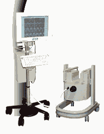
From MagneVu;
The MagneVu 1000 is a compact, robust, and portable, permanent magnet MRI system and operates without special shielding or costly site preparation.
This MRI device utilizes a patented non-homogeneous magnetic field image acquisition method to achieve high performance imaging. The MagneVu 1000 MRI scanner is designed for MRI of the extremities with the current specialty areas in diabetes and rheumatoid arthritis. Easy access is afforded for claustrophobic, pediatric, or limited mobility patients. In August 1998
FDA marketing clearance and other regulatory approvals have been received. Until 2008, over 130 devices in the US are in use. Some further developments of MagneVu's extremity scanner are: 'truly Plug n' Play MRI™' and iSiS ( which adds wireless capability to the second generation MV1000-XL).
Device Information and Specification IMAGING MODES 3-dimensional multi-echo data acquisition | |  | |
• View the DATABASE results for 'MagneVu 1000' (3).
| | | | |  Further Reading: Further Reading: | News & More:
|
|
| |
|  | |  |  |
|  | | |
|
| |
 | Look
Ups |
| |