Magnetic Resonance - Technology Information Portal Welcome to MRI Technology
Info
  Sheets

Out-
      side


 'T2 Weighted Image' 
SEARCH FOR    
 
  2 3 5 A B C D E F G H I J K L M N O P Q R S T U V W X Y Z
Result : Searchterm 'T2 Weighted Image' found in 1 term [] and 19 definitions []
1 - 5 (of 20)     next
Result Pages : [1]  [2 3 4]
Searchterm 'T2 Weighted Image' was also found in the following services: 
spacer
News  (4)  Resources  (1)  
 
T2 Weighted ImageInfoSheet: - Sequences - 
Intro, 
Overview, 
Types of, 
etc.
 
T2 weighted imaging relies upon local dephasing of spins following the application of the transverse energy pulse. The contrast of a T2 weighted image is predominantly dependent on T2 and the T2 dependence will be increased by using a long echo time.
Fat has a shorter T2 time than water and relaxes or decays more readily than water. Since the amount of transverse magnetization in fat is small, fat generates very little signal on a strong T2 weighted contrast image and appears intermediate to dark. The T2 weighting is stronger with a longer TE. Water has a very high T2 constant, therefore has very high T2 signal and thus appears bright on a T2 contrast image. Cerebral white matter (fat containing) is less intense than grey matter. Flowing blood (flow effects) and haematomas (haemoglobin, haemosiderin) have a variable signal intensity on MR images.
Images created with TR's and TE's to enhance T2 contrast are referred to as T2 weighted images. Both T1 and T2 weighted images are acquired for most medical MRI examinations.
 
Images, Movies, Sliders:
 Lumbar Spine T2 FSE Sagittal  Open this link in a new window
    

Courtesy of  Robert R. Edelman
 Shoulder Axial T2 FatSat FRFSE  Open this link in a new window
    

Courtesy of  Robert R. Edelman
 Lumbar Spine T2 FSE Axial  Open this link in a new window
 
spacer
 
• Share the entry 'T2 Weighted Image':  Facebook  Twitter  LinkedIn  
 
• Related Searches:
    • Brain MRI
    • MRI Procedure
    • T2*
    • Breast MRI
    • Relaxation Effect
 
Further Reading:
  Basics:
MYELIN-SELECTIVE MRI: PULSE SEQUENCE DESIGN AND OPTIMIZATION
   by www.imaging.robarts.ca    
T2* cardiac MRI allows prediction of severe reperfusion injury after STEMI
Tuesday, 9 November 2010   by www.medwire-news.md    
  News & More:
Periodical assessment of four horns of knee meniscus using MR T2 mapping imaging in volunteers before and after amateur marathons
Friday, 15 July 2022   by www.nature.com    
New MRI technique offers faster diagnosis of multiple sclerosis
Monday, 1 February 2016   by medicalxpress.com    
MRI measurement tools to help diagnose veterans' traumatic brain injuries
Wednesday, 18 March 2015   by medicalxpress.com    
New MR sequence helps radiologists more accurately evaluate abnormalities of the uterus and ovaries
Thursday, 23 April 2009   by www.eurekalert.org    
MRI Resources 
Breast MRI - Education - Distributors - Patient Information - Diffusion Weighted Imaging - Databases
 
ContrastForum -
related threads
 
Contrast is the relative difference of signal intensities in two adjacent regions of an image.
Due to the T1 and T2 relaxation properties in magnetic resonance imaging, differentiation between various tissues in the body is possible. Tissue contrast is affected by not only the T1 and T2 values of specific tissues, but also the differences in the magnetic field strength, temperature changes, and many other factors. Good tissue contrast relies on optimal selection of appropriate pulse sequences (spin echo, inversion recovery, gradient echo, turbo sequences and slice profile).
Important pulse sequence parameters are TR (repetition time), TE (time to echo or echo time), TI (time for inversion or inversion time) and flip angle. They are associated with such parameters as proton density and T1 or T2 relaxation times. The values of these parameters are influenced differently by different tissues and by healthy and diseased sections of the same tissue.
For the T1 weighting it is important to select a correct TR or TI. T2 weighted images depend on a correct choice of the TE. Tissues vary in their T1 and T2 times, which are manipulated in MRI by selection of TR, TI, and TE, respectively. Flip angles mainly affect the strength of the signal measured, but also affect the TR/TI/TE parameters.
Conditions necessary to produce different weighted images:
T1 Weighted Image: TR value equal or less than the tissue specific T1 time - TE value less than the tissue specific T2 time.
T2 Weighted Image: TR value much greater than the tissue specific T1 time - TE value greater or equal than the tissue specific T2 time.
Proton Density Weighted Image: TR value much greater than the tissue specific T1 time - TE value less than the tissue specific T2 time.

See also Image Contrast Characteristics, Contrast Reversal, Contrast Resolution, and Contrast to Noise Ratio.
 
Images, Movies, Sliders:
 Fetus (Brain) and Dermoid in Mother  Open this link in a new window
      

Courtesy of  Robert R. Edelman

 Circle of Willis, Time of Flight, MIP  Open this link in a new window
    
SlidersSliders Overview

 Anatomic MRI of the Knee 1  Open this link in a new window
    
SlidersSliders Overview

 Anatomic Imaging of the Liver  Open this link in a new window
      

 Brain MRI Inversion Recovery  Open this link in a new window
    
 
spacer

• View the DATABASE results for 'Contrast' (373).Open this link in a new window


• View the NEWS results for 'Contrast' (77).Open this link in a new window.
 
Further Reading:
  Basics:
Magnetic resonance imaging
   by www.scholarpedia.org    
MRI's inside story
Thursday, 4 December 2003   by www.economist.com    
Image Characteristics and Quality
   by www.sprawls.org    
  News & More:
A natural boost for MRI scans
Monday, 21 October 2013   by www.eurekalert.org    
A groundbreaking new graphene-based MRI contrast agent
Friday, 8 June 2012   by www.nanowerk.com    
New MRI Chemical Offers Amazing Contrast
Friday, 22 January 2010   by news.softpedia.com    
MRI Resources 
Spectroscopy - Pacemaker - MR Myelography - Implant and Prosthesis - Education pool - DICOM
 
Fast Spin EchoForum -
related threadsInfoSheet: - Sequences - 
Intro, 
Overview, 
Types of, 
etc.
 
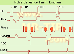
Fast Spin Echo Diagram (FSE) In the pulse sequence timing diagram, a fast spin echo sequence with an echo train length of 3 is illustrated. This sequence is characterized by a series of rapidly applied 180° rephasing pulses and multiple echoes, changing the phase encoding gradient for each echo.
The echo time TE may vary from echo to echo in the echo train. The echoes in the center of the K-space (in the case of linear k-space acquisition) mainly produce the type of image contrast, whereas the periphery of K-space determines the spatial resolution. For example, in the middle of K-space the late echoes of T2 weighted images are encoded. T1 or PD contrast is produced from the early echoes.
The benefit of this technique is that the scan duration with, e.g. a turbo spin echo turbo factor / echo train length of 9, is one ninth of the time. In T1 weighted and proton density weighted sequences, there is a limit to how large the ETL can be (e.g. a usual ETL for T1 weighted images is between 3 and 7). The use of large echo train lengths with short TE results in blurring and loss of contrast. For this reason, T2 weighted imaging profits most from this technique.
In T2 weighted FSE images, both water and fat are hyperintense. This is because the succession of 180° RF pulses reduces the spin spin interactions in fat and increases its T2 decay time. Fast spin echo (FSE) sequences have replaced conventional T2 weighted spin echo sequences for most clinical applications. Fast spin echo allows reduced acquisition times and enables T2 weighted breath hold imaging, e.g. for applications in the upper abdomen.
In case of the acquisition of 2 echoes this type of a sequence is named double fast spin echo / dual echo sequence, the first echo is usually density and the second echo is T2 weighted image. Fast spin echo images are more T2 weighted, which makes it difficult to obtain true proton density weighted images. For dual echo imaging with density weighting, the TR should be kept between 2000 - 2400 msec with a short ETL (e.g., 4).
Other terms for this technique are:
Turbo Spin Echo
Rapid Imaging Spin Echo,
Rapid Spin Echo,
Rapid Acquisition Spin Echo,
Rapid Acquisition with Refocused Echoes
 
Images, Movies, Sliders:
 Lumbar Spine T2 FSE Sagittal  Open this link in a new window
    

Courtesy of  Robert R. Edelman
 MRI - Anatomic Imaging of the Foot  Open this link in a new window
    
SlidersSliders Overview

 Lumbar Spine T2 FSE Axial  Open this link in a new window
    

Courtesy of  Robert R. Edelman
 
spacer

• View the DATABASE results for 'Fast Spin Echo' (31).Open this link in a new window

 
Further Reading:
  Basics:
MYELIN-SELECTIVE MRI: PULSE SEQUENCE DESIGN AND OPTIMIZATION
   by www.imaging.robarts.ca    
Advances in Magnetic Resonance Neuroimaging
Friday, 27 February 2009   by www.ncbi.nlm.nih.gov    
  News & More:
New MR sequence helps radiologists more accurately evaluate abnormalities of the uterus and ovaries
Thursday, 23 April 2009   by www.eurekalert.org    
Spin echoes, CPMG and T2 relaxation - Introductory NMR & MRI from Magritek
2013   by www.azom.com    
Searchterm 'T2 Weighted Image' was also found in the following services: 
spacer
News  (4)  Resources  (1)  
 
Signal Intensity
 
Signal intensity interpretation in MR imaging has a major problem.
Often there is no intuitive approach to signal behavior as signal intensity is a very complicated function of the contrast-determining tissue parameter, proton density, T1 and T2, and the machine parameters TR and TE. For this reason, the terms T1 weighted image, T2 weighted image and proton density weighted image were introduced into clinical MR imaging.
Air and bone produce low-intensity, weaker signals with darker images. Fat and marrow produce high-intensity signals with brighter images.
The signal intensity measured is related to the square of the xy-magnetization, which in a SE pulse sequence is given by
Mxy = Mxy0(1-exp(-TR/T1)) exp(-TE/T2) (1)
where Mxy0 = Mz0 is proportional to the proton or spin density, and corresponds to the z-magnetization present at zero time of the experiment when it is tilted into the xy-plane.

See also T2 Weighted Image and Ernst Angle.
spacer

• View the DATABASE results for 'Signal Intensity' (56).Open this link in a new window


• View the NEWS results for 'Signal Intensity' (1).Open this link in a new window.
 
Further Reading:
  Basics:
Contrast mechanisms in magnetic resonance imaging
2004   by www.iop.org    
Image Characteristics and Quality
   by www.sprawls.org    
  News & More:
Specialized MRI sensor can detect light deep within tissues
Thursday, 22 December 2022
Study compares effect of food intake on manganese-based MRI contrast agent absorption
Saturday, 3 December 2022   by www.itnonline.com    
Kidney stone imaging with 3D ultra-short echo time (UTE) magnetic resonance imaging. A phantom study - Abstract
Wednesday, 11 March 2015   by www.urotoday.com    
MRI Resources 
Online Books - MRI Technician and Technologist Schools - PACS - Corporations - Spectroscopy pool - MRI Physics
 
T2 TimeForum -
related threads
 
The T2 relaxation time (spin spin relaxation time or transverse relaxation time), is a biological parameter that is used in MRIs to distinguish between tissue types and is termed 'Time 2' or T2. It is a tissue-specific time constant for protons and is dependent on the exchanging of energy with near by nuclei. T2 weighted images rely upon local dephasing of spins following the application of the transverse energy pulse. T2 is the decay of magnetization perpendicular to the main magnetic field (in an ideal homogeneous field).
Due to interaction between the spins, they lose their phase coherence, which results in a loss of transverse magnetization and MRI signal. After time T2 transverse magnetization has lost 63% of its original value. This tissue parameter determines the contrast.
The T2 relaxation is temperature dependent. At a lower temperature molecular motion is reduced and the decay times are reduced.
Fat has a very efficient energy exchange and therefore it has a relatively short T2.
Water is less efficient than fat in the exchange of energy, and therefore it has a long T2 time.

See also T2 Weighted Image and Magnetic Resonance Imaging MRI.
 
Images, Movies, Sliders:
 Breast MRI Images T2 And T1  Open this link in a new window
      
 MRI of the Skull Base  Open this link in a new window
    
SlidersSliders Overview

 Knee MRI Transverse 002  Open this link in a new window
    
 
spacer

• View the DATABASE results for 'T2 Time' (16).Open this link in a new window

 
Further Reading:
  News & More:
MYELIN-SELECTIVE MRI: PULSE SEQUENCE DESIGN AND OPTIMIZATION
   by www.imaging.robarts.ca    
MRI Resources 
MR Guided Interventions - General - Pacemaker - Contrast Agents - Cardiovascular Imaging - Nerve Stimulator
 
     1 - 5 (of 20)     next
Result Pages : [1]  [2 3 4]
 Random Page
 
Share This Page
FacebookTwitterLinkedIn

MR-TIP    
Community   
User
Pass
Forgot your UserID/Password ?    


Next big thing in MRI will be :
AI 
remote operator 
personalized protocols 
helium-free 
molecular MRI 
portable MRI 

Look
      Ups


MR-TIP.com uses cookies! By browsing MR-TIP.com, you agree to our use of cookies.

Magnetic Resonance - Technology Information Portal
Member of SoftWays' Medical Imaging Group - MR-TIP • Radiology-TIP • Medical-Ultrasound-Imaging • 
Copyright © 2003 - 2024 SoftWays. All rights reserved. [ 18 April 2024]
Terms of Use | Privacy Policy | Advertising
 [last update: 2024-02-26 03:41:00]