| Info
Sheets |
| | | | | | | | | | | | | | | | | | | | | | | | |
 | Out-
side |
| | | | |
|
| | | | | |  | Searchterm 'SPIR' was also found in the following services: | | | | |
|  |  |
| |
|
From Siemens Medical Systems;
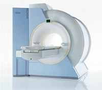
while older navigator techniques take up to 40 minutes to create, the high performance of the MAGNETOM Sonata system enables 'complete examinations in less than 15 minutes'. It creates a new standard of diagnostic confidence and moves Cardiac MR from the research setting into routine clinical practice.
Device Information and Specification CLINICAL APPLICATION Whole body Body, head, spine, knee, neck, TMJ, extremity, head, breast, shoulder, others GRE, IR, FIR, STIR, TrueIR/FISP, FSE, FLAIR, MT, SS-FSE, MT-SE, MTC, MSE, EPI, 3D DESS//CISS/PSIF, GMR IMAGING MODES Single, multislice, volume study, multi angle, multi oblique178 images/sec at 256 x 256 at 100% FOV1024 x 1024 full screen display 4050kg, 5500kg in operation POWER REQUIREMENTS 380/400/420/440/480 V Passive, act.; 1st order std./2nd opt. | | | | | | | | | | |
| | | | | |
| |
|
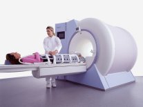
Device Information and Specification CLINICAL APPLICATION Whole body GRE, IR, FIR, STIR, TrueIR/FISP, FSE, FLAIR, MT, SS-FSE, MT-SE, MTC, MSE, EPI, GMR, fat/water sat./exc. IMAGING MODES Single, multislice, volume study, multi angle, multi obliqueTR 2.4 msec std.; 2.0 opt.; 1.8 w/30 mT/m at 256matrix TE 1.1 msec std.; 0.9 opt.; 0.78 w/30 mT/m at 256matrix 178 images/sec at 256 x 256 at 100% FOV1024 x 1024 full screen display 21 micrometer in plane, 11 micrometer optional 4050kg, 5500kg in operation H*W*D 236 x 215 x 160 cm w/covers POWER REQUIREMENTS 380/400/420/440/480 V STRENGTH 20/35 mT/m standard, 30/52 opt. Passive, act.; 1st order std./2nd opt. | | | |
• View the DATABASE results for 'MAGNETOM Symphony™' (2).
| | | | |  Further Reading: Further Reading: | Basics:
|
|
| |
| | | | | |
| |
|
From Siemens Medical Systems;
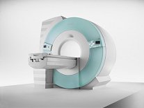
70 cm + 125 cm + 1.5T and Tim - a combination never seen before in MRI ...
MAGNETOM Espree™s unique open bore design can accommodate more types of patients than other 1.5T systems on the market today, in particular the growing population of obese patients. The power of 1.5T combined with Tim technology boosts signal to noise, which is necessary to adequately image obese patients.
Device Information and Specification
CLINICAL APPLICATION
Whole body
Body, Tim [32 x 8], Tim [76 coil elements with up to 18 RF channels])
GRE, IR, FIR, STIR, TrueIR/FISP, FSE, FLAIR, MT, SS-FSE, MT-SE, MTC, MSE, EPI, 3D DESS//CISS/PSIF, GMR
IMAGING MODES
Single, multislice, volume study, multi angle, multi oblique
Image Processor reconstructing up to 3226 images per second (256 x 256, 25% recFoV)
1024 x 1024 full screen display
| | | |
• View the DATABASE results for 'MAGNETOM Espree™' (2).
| | | | | | Further Reading: | News & More:
|
|
| |
| | | Searchterm 'SPIR' was also found in the following services: | | | | |
| | |
| |
|
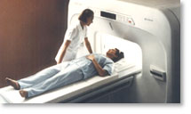
From Hitachi Medical Systems America, Inc.; because of its dependability, the MRP-7000™ remains popular more than a decade after the first U.S. system was shipped. This system maintains a high resale value, what has made it one of the most sought-after scanners on the used MRI equipment market.
Device Information and Specification CLINICAL APPLICATION Whole body DualQuad T/R Body Coil, MA Head, MA C-Spine, MA Shoulder, MA Wrist, MA CTL Spine, MA Knee, MA TMJ, MA Flex Body (3 sizes), Neck, small and large Extremity, PVA (WIP), Breast (WIP), Neurovascular (WIP), Cardiac (WIP) and MA Foot//Ankle (WIP) SE, GE, GR, IR, FIR, STIR, ss-FSE, FSE, DE-FSE/FIR, FLAIR, ss/ms-EPI, ss/ms EPI- DWI, SSP, MTC, SE/GE-EPI, MRCP, SARGE, RSSG, TRSG, BASG, Angiography: CE, PC, 2D/3D TOFIMAGING MODES Single, multislice, volume study horizontal 2.5 m x 2.1 m vertical | | | |
• View the DATABASE results for 'MRP-7000™' (2).
| | | | |
| | | | | |
| |
|
( MRI) Magnetic resonance imaging is a noninvasive medical imaging technique that uses the interaction between radio frequency pulses, a strong magnetic field and body tissue to obtain images of slices/planes from inside the body. These magnets generate fields from approx. 2000 times up to 30000 times stronger than that of the Earth. The use of nuclear magnetic resonance principles produces extremely detailed pictures of the body tissue without the need for x-ray exposure and gives diagnostic information of various organs.
Measured are mobile hydrogen nuclei (protons are the hydrogen atoms of water, the 'H' in H 20), the majority of elements in the body. Only a small part of them contribute to the measured signal, caused by their different alignment in the magnetic field. Protons are capable of absorbing energy if exposed to short radio wave pulses (electromagnetic energy) at their resonance frequency. After the absorption of this energy, the nuclei release this energy so that they return to their initial state of equilibrium.
This transmission of energy by the nuclei as they return to their initial state is what is observed as the MRI signal. The subtle differing characteristic of that signal from different tissues combined with complex mathematical formulas analyzed on modern computers is what enables MRI imaging to distinguish between various organs. Any imaging plane, or slice, can be projected, and then stored or printed.
The measured signal intensity depends jointly on the spin density and the relaxation times ( T1 time and T2 time), with their relative importance depending on the particular imaging technique and choice of interpulse times. Any motion such as blood flow, re spiration, etc. also affects the image brightness.
Magnetic resonance imaging is particularly sensitive in assessing anatomical structures, organs and soft tissues for the detection and diagnosis of a broad range of pathological conditions. MRI pictures can provide contrast between benign and pathological tissues and may be used to stage cancers as well as to evaluate the response to treatment of malignancies. The need for biopsy or exploratory surgery can be eliminated in some cases, and can result in earlier diagnosis of many diseases. See also MRI History and Functional Magnetic Resonance Imaging (fMRI). | | | | | |
• View the DATABASE results for 'Magnetic Resonance Imaging MRI' (9).
| | |
• View the NEWS results for 'Magnetic Resonance Imaging MRI' (222).
| | | | | | Further Reading: | | Basics:
|
|
News & More:
| |
| |
| | | | |
| | |
| | | |
|
| |
 | Look
Ups |
| |