Magnetic Resonance - Technology Information Portal Welcome to MRI Technology
Info
  Sheets

Out-
      side


 'SNR' 
SEARCH FOR    
 
  2 3 5 A B C D E F G H I J K L M N O P Q R S T U V W X Y Z
Result : Searchterm 'SNR' found in 0 term [] and 46 definitions []
previous     11 - 15 (of 46)     next
Result Pages : [1 2 3 4 5 6 7 8 9 10]
Searchterm 'SNR' was also found in the following services: 
spacer
News  (3)  Forum  (11)  
 
Imaging CoilInfoSheet: - Coils - 
Intro, 
Overview, 
etc.MRI Resource Directory:
 - Coils -
 
Imaging coils are radio frequency coils used in magnetic resonance imaging for sending and/or receiving electromagnetic radiation. Several MR imaging coils in different shapes and sizes are necessary for different body parts and to handle individual applications.
For Example:
Birdcage coils provide high homogeneity and good signal to noise ratio (SNR) in brain MRI scans.
Surface coils with small coil diameter and higher SNR enable to image the temporomandibular joints (TMJ).
The implemented body coil allows the scanning of body parts with large field of views.
Micro coils are available to image finger joints.
High performance phased array coils are today state-of-the-art for a wide range of applications from head, spine, knee or shoulder to cardiac MRI.
For different types of coils see Volume Coil, Sense Coil, Array Coil, Surface Coil, and Bird Cage Coil.

See also the related poll result: '3rd party coils are better than the original manufacturer coils'
spacer

• View the NEWS results for 'Imaging Coil' (9).Open this link in a new window.
 
Further Reading:
  Basics:
Radio-frequency Coil Selection for MR Imaging of the Brain and Skull Base1
   by radiology.rsnajnls.org    
MRI Resources 
MRI Technician and Technologist Career - Collections - Research Labs - Brain MRI - Sequences - Services and Supplies
 
MAGNETOM Aera
 
www.healthcare.siemens.com/magnetic-resonance-imaging/0-35-to-1-5t-mri-scanner/magnetom-aera/ 
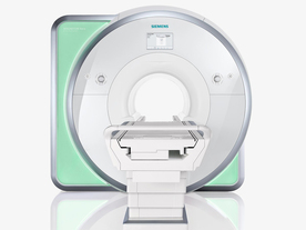
From Siemens Medical Systems;
Received FDA clearance in 2010.
The MAGNETOM Aera is a patient friendly, comfortable 1.5 Tesla MRI system with advanced radio frequency chain.
The system is equipped with the Tim 4G and Dot system (Total imaging matrix + Day optimizing throughput), to enhance both productivity and image quality.
Tim 4G technology provides improved SNR. The standard system configuration of 48 radio frequency channels and 204 coil elements creates an imaging matrix that allows maximum use of coil elements at full field of view. Dot provides improved image consistency through new features like auto align, auto FoV and automatic bolus detection.
Device Information and Specification
CLINICAL APPLICATION
Whole body
CONFIGURATION
Open bore
Head, spine, torso/ body coil, neurovascular, cardiac, neck, shoulder, knee, wrist, foot//ankle and multi-purpose flex coils. Peripheral vascular, breast, shoulder. Up to 60% more SNR with Tim 4G.
CHANNELS (min. / max. configuration)
48, 64
IMAGING TECHNIQUES
iPAT, mSENSE and GRAPPA (image, k-space), noncontrast angiography, plaque imaging, radial motion compensation, Dixon, improved workflow with Dot, Caipirinha - single digit breath-holds for 3-D body imaging.
MINIMUM TR
3-D GRE: 0.95 (256 matrix)
MINIMUM TE
3-D GRE: 0.22 (256 matrix), Ultra-short TE
FOV
0.5 - 50
BORE DIAMETER
or W x H
At isocenter: L-R 70 cm, A-P (with table) 55 cm
TABLE CAPACITY
250 kg
MAGNET WEIGHT (gantry included)
3121 kg
DIMENSION H*W*D (gantry included)
145 x 231 x 219 cm
5-GAUSS FRINGE FIELD
2.5 m / 4.0 m
CRYOGEN USE
Zero boil off rate, approx. 10 years
COOLING SYSTEM
Water
up to 200 T/m/s
MAX. AMPLITUDE
33 or 45 mT/m
3 linear with 20 coils, 5 nonlinear 2nd-order
POWER REQUIREMENTS
380 / 400 / 420 / 440 / 460 / 480 V, 3-phase + ground; 85 kVA
spacer
MRI Resources 
Homepages - Jobs pool - Cochlear Implant - Used and Refurbished MRI Equipment - Pacemaker - Sequences
 
MEDIC TechniqueInfoSheet: - Sequences - 
Intro, 
Overview, 
Types of, 
etc.
 
Multiple echoes acquired in a gradient echo scan are combined into an image, for less artifacts and higher SNR; the early echoes provide increased SNR, whilst later echoes boost contrast. This sequence type is also known as MERGE (Multiple Echo Recombined Gradient Echo)

See also Multi Echo Data Image Combination, Bipolar Gradient Pulse.
spacer

• View the NEWS results for 'MEDIC Technique' (15).Open this link in a new window.
 
Further Reading:
  Basics:
Cutting Edge Imaging of THE Spine
February 2007   by www.pubmedcentral.nih.gov    
Searchterm 'SNR' was also found in the following services: 
spacer
News  (3)  Forum  (11)  
 
Matrix Size
 
The number of data points collected in one, two or all three directions. Normally used for the 2D in plane sampling. The display matrix may be different from the acquisition matrix, although the latter determines the resolution. Measurement time may be saved by not acquiring raw data lines corresponding to high resolution. Not measured rows are filled with zeroes prior to the image calculation. A square image is the result of an interpolation in phase encoding direction. See also Zero Filling.
mri safety guidance
Image Guidance
The chosen matrix size effects scan time, resolution and SNR. Reduced measurement matrixes decrease the scan time and the resolution by increased SNR.
spacer

• View the DATABASE results for 'Matrix Size' (4).Open this link in a new window

MRI Resources 
MRI Reimbursement - MRI Centers - Pacemaker - Online Books - PACS - MRI Technician and Technologist Jobs
 
Partial Echo
 
(PE) The partial echo technique (also called fractional echo) is used to shorten the minimum echo time. By the acquisition of only a part of k-space data this technique benefits (like all partial Fourier techniques) from the complex conjugate symmetry between the k-space halves (this is called Hermitian symmetry).
The dephasing gradient in the frequency direction is reduced, and the duration of the readout gradient and the data acquisition window are shortened. Partial echo gives a better SNR at a given TE when a smaller FOV or thinner slices are selected, allows a longer sampling time, and a larger water fat shift (WFS, see also bandwidth) due to a lower gradient amplitude. The resolution is not affected. This is often used in gradient echo sequences (e.g. FLASH, Contrast Enhanced Magnetic Resonance Angiography) to reduce the echo time and yields a lower gradient moment. The disadvantage of using a partial echo can be a lower SNR, although this may be partly offset by the reduced echo time.
Also called Fractional Echo, Read Conjugate Symmetry, Single Side View.

See also Partial Fourier Technique and acronyms for 'partial echo' from different manufacturers.
spacer

• View the DATABASE results for 'Partial Echo' (4).Open this link in a new window

 
Further Reading:
  Basics:
Method and apparatus for subterranean formation flow imaging
   by www.google.com    
MRI Resources 
Spectroscopy - Blood Flow Imaging - Sequences - Open Directory Project - Developers - Guidance
 
previous      11 - 15 (of 46)     next
Result Pages : [1 2 3 4 5 6 7 8 9 10]
 Random Page
 
Share This Page
FacebookTwitterLinkedIn

MR-TIP    
Community   
User
Pass
Forgot your UserID/Password ?    


MRI is trending to low field magnets :
reduced costs will lead to this change 
AI will close the gap to high field 
only in remote areas 
is only temporary 
never 

Look
      Ups


MR-TIP.com uses cookies! By browsing MR-TIP.com, you agree to our use of cookies.

Magnetic Resonance - Technology Information Portal
Member of SoftWays' Medical Imaging Group - MR-TIP • Radiology-TIP • Medical-Ultrasound-Imaging • 
Copyright © 2003 - 2024 SoftWays. All rights reserved. [ 26 April 2024]
Terms of Use | Privacy Policy | Advertising
 [last update: 2024-02-26 03:41:00]