 | Info
Sheets |
| | | | | | | | | | | | | | | | | | | | | | | | |
 | Out-
side |
| | | | |
|
| | | | | |  | Searchterm 'Meter' was also found in the following services: | | | | |
|  |  |
| |
|
(IR) Inversion recovery is an MRI technique, which can be incorporated into MR imaging, wherein the nuclear magnetization is inverted at a time on the order of T1 before the regular imaging pulse-gradient sequences. The resulting partial relaxation of the spins in the different structures being imaged can be used to produce an image that depends strongly on T1. This may bring out differences in the appearance of structures with different T1 relaxation times. Note that this does not directly produce an image of T1. T1 in a given region can be calculated from the change in the MR signal from the region due to the inversion pulse compared to the signal with no inversion pulse or an inversion pulse with a different inversion time. This sequence involves successive 180° and 90° pulses. The inversion recovery sequence is specified in terms of three para meters, inversion time (TI), repetition time (TR) and echo time (TE). See also Inversion Recovery Sequence and FLAIR. | | | | | | | | | | | | |  Further Reading: Further Reading: | | Basics:
|
|
News & More:
| |
| |
| | | Searchterm 'Meter' was also found in the following services: | | | | |
| | |
| |
|
| | | | | |
• View the DATABASE results for 'Inversion Time' (14).
| | | | | | Further Reading: | News & More:
|
|
| |
| | | | | |
| |
|
(J) The SI unit of work or energy.
Definition: The work done by a force of 1 newton acting to move an object through a distance of 1 meter in the direction in which the force is applied.
Since kinetic energy is one half the mass times the square of the velocity, 1 joule is the kinetic energy of a mass of two kilograms moving at a velocity of 1 m/sec.
The joule is named for the British physicist James P. Joule. | | | |
• View the DATABASE results for 'Joule' (5).
| | | | |
| | | Searchterm 'Meter' was also found in the following services: | | | | |
| | |
| |
|
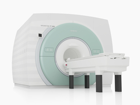
From Siemens Medical Systems;
The MAGNETOM 7T is designed as an open research platform. 7T MRI provides anatomical detail at the submillimiter scale, enhanced contrast mechanisms, outstanding spectroscopy performance, ultra-high resolution functional imaging ( fMRI), multinuclear whole-body MRI and functional information.
This ultra high field (UHF) MRI device is a research system and not cleared, approved or licensed in any jurisdiction for patient examinations.
Device Information and Specification
CLINICAL APPLICATION
Whole body
High-performance, ultra high field coils available. Integration and support for coil developments.
CHANNELS (min. / max. configuration)
32, optional 8 channels TX array
40 x 40 x 30 cm³ less than 8% nonlinearity
MAGNET WEIGHT (gantry included)
35017 kg
DIMENSION H*W*D (gantry included)
320 x 240 x 317,5 cm
MAX. AMPLITUDE
up to 70 mT/m
Up to 3rd order shim coils, user configurable B0 shim ? B0 maps and ROI definition
POWER REQUIREMENTS
2000 Volts, 650A
| | | | | | | Further Reading: | Basics:
|
|
News & More:
| |
| |
| | | Searchterm 'Meter' was also found in the following services: | | | | |
| | |
| |
|
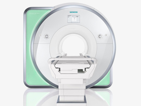
From Siemens Medical Systems;
Received FDA clearance in 2010.
The MAGNETOM Aera is a patient friendly, comfortable 1.5 Tesla MRI system with advanced radio frequency chain.
The system is equipped with the Tim 4G and Dot system (Total imaging matrix + Day optimizing throughput), to enhance both productivity and image quality.
Tim 4G technology provides improved SNR. The standard system configuration of 48 radio frequency channels and 204 coil elements creates an imaging matrix that allows maximum use of coil elements at full field of view. Dot provides improved image consistency through new features like auto align, auto FoV and automatic bolus detection.
Device Information and Specification
CLINICAL APPLICATION
Whole body
Head, spine, torso/ body coil, neurovascular, cardiac, neck, shoulder, knee, wrist, foot//ankle and multi-purpose flex coils. Peripheral vascular, breast, shoulder. Up to 60% more SNR with Tim 4G.
CHANNELS (min. / max. configuration)
48, 64
MINIMUM TE
3-D GRE: 0.22 (256 matrix), Ultra-short TE
At isocenter: L-R 70 cm, A-P (with table) 55 cm
MAGNET WEIGHT (gantry included)
3121 kg
DIMENSION H*W*D (gantry included)
145 x 231 x 219 cm
MAX. AMPLITUDE
33 or 45 mT/m
3 linear with 20 coils, 5 nonlinear 2nd-order
POWER REQUIREMENTS
380 / 400 / 420 / 440 / 460 / 480 V, 3-phase + ground; 85 kVA
| | | | | |
| | | | |
| | | |
|
| |
 | Look
Ups |
| |


