 | Info
Sheets |
| | | | | | | | | | | | | | | | | | | | | | | | |
 | Out-
side |
| | | | |
|
| | | | | |  | Searchterm 'Gauss' was also found in the following services: | | | | |
|  |  |
| |
|
| | | | | | | | | | |  Further Reading: Further Reading: | | Basics:
|
|
News & More:
|  |
Safety of Bedside Portable Low-Field Brain MRI in ECMO Patients Supported on Intra-Aortic Balloon Pump
Friday, 18 November 2022 by www.mdpi.com | | |
Researchers at the University of Tsukuba develop a portable MRI system specifically for identifying wrist cartilage damage among athletes, providing a convenient means of early detection and treatment of injuries
Tuesday, 26 April 2022 by www.tsukuba.ac.jp | | |
This bizarre looking helmet can create better brain scans
Friday, 11 February 2022 by www.sciencedaily.com | | |
A low-cost and shielding-free ultra-low-field brain MRI scanner
Tuesday, 14 December 2021 by www.nature.com | | |
Portable MRI provides life-saving information to doctors treating strokes
Thursday, 5 August 2021 by news.yale.edu | | |
Synaptive Evry, an MRI for Any Space, Cleared by FDA
Thursday, 30 April 2020 by www.medgadget.com | | |
World's First Portable MRI Cleared by FDA
Monday, 17 February 2020 by www.medgadget.com | | |
Introducing a point-of-care MRI system
Tuesday, 29 October 2019 by healthcare-in-europe.com | | |
Opportunities in Interventional and Diagnostic Imaging by Using High-performance Low-Field-Strength MRI
Tuesday, 1 October 2019 by pubs.rsna.org | | |
Portable 'battlefield MRI' comes out of the lab
Thursday, 30 April 2015 by physicsworld.com | | |
Portable MRI could aid wounded soldiers and children in the third world
Thursday, 23 April 2015 by phys.org |
|
| |
| | | Searchterm 'Gauss' was also found in the following service: | | | | |
| | |
| |
|
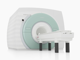
From Siemens Medical Systems;
The MAGNETOM 7T is designed as an open research platform. 7T MRI provides anatomical detail at the submillimiter scale, enhanced contrast mechanisms, outstanding spectroscopy performance, ultra-high resolution functional imaging ( fMRI), multinuclear whole-body MRI and functional information.
This ultra high field (UHF) MRI device is a research system and not cleared, approved or licensed in any jurisdiction for patient examinations.
Device Information and Specification
CLINICAL APPLICATION
Whole body
High-performance, ultra high field coils available. Integration and support for coil developments.
CHANNELS (min. / max. configuration)
32, optional 8 channels TX array
40 x 40 x 30 cm³ less than 8% nonlinearity
MAGNET WEIGHT (gantry included)
35017 kg
DIMENSION H*W*D (gantry included)
320 x 240 x 317,5 cm
MAX. AMPLITUDE
up to 70 mT/m
Up to 3rd order shim coils, user configurable B0 shim ? B0 maps and ROI definition
POWER REQUIREMENTS
2000 Volts, 650A
| | | | | | | Further Reading: | Basics:
|
|
News & More:
| |
| |
| | | | | |
| |
|
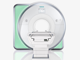
From Siemens Medical Systems;
Received FDA clearance in 2010.
The MAGNETOM Aera is a patient friendly, comfortable 1.5 Tesla MRI system with advanced radio frequency chain.
The system is equipped with the Tim 4G and Dot system (Total imaging matrix + Day optimizing throughput), to enhance both productivity and image quality.
Tim 4G technology provides improved SNR. The standard system configuration of 48 radio frequency channels and 204 coil elements creates an imaging matrix that allows maximum use of coil elements at full field of view. Dot provides improved image consistency through new features like auto align, auto FoV and automatic bolus detection.
Device Information and Specification
CLINICAL APPLICATION
Whole body
Head, spine, torso/ body coil, neurovascular, cardiac, neck, shoulder, knee, wrist, foot//ankle and multi-purpose flex coils. Peripheral vascular, breast, shoulder. Up to 60% more SNR with Tim 4G.
CHANNELS (min. / max. configuration)
48, 64
MINIMUM TE
3-D GRE: 0.22 (256 matrix), Ultra-short TE
At isocenter: L-R 70 cm, A-P (with table) 55 cm
MAGNET WEIGHT (gantry included)
3121 kg
DIMENSION H*W*D (gantry included)
145 x 231 x 219 cm
MAX. AMPLITUDE
33 or 45 mT/m
3 linear with 20 coils, 5 nonlinear 2nd-order
POWER REQUIREMENTS
380 / 400 / 420 / 440 / 460 / 480 V, 3-phase + ground; 85 kVA
| | | | | |
| | | Searchterm 'Gauss' was also found in the following services: | | | | |
| | |
| |
|
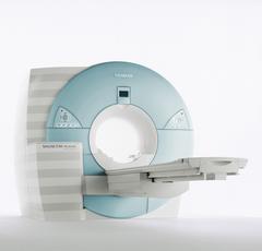
From Siemens Medical Systems;
MAGNETOM Avanto with Tim - Total imaging matrix technology.
For true whole-body anatomical coverage. For ultra-fast image
acquisition. Aids the physician in fast and precise
evaluation of systemic diseases like colon cancer, metastasis imaging, vessel diseases, and preventional exams. For claustrophobic patients,
MAGNETOM Avanto enables feetfirst exams for nearly all MR procedures. For obese patients, MAGNETOM Avanto supports up to 200 kg (400 lbs), without table movement restrictions. The AudioComfort technology enables up to a 30 dB(A) acoustic noise reduction, that means nearly all clinical routine sequences are running under 99 dB(A).
Device Information and Specification
CLINICAL APPLICATION
Whole body
CONFIGURATION
Compact short bore
Body, Tim [32 x 8], Tim [76 x18], Tim [76 coil elements with up to 32 RF channels]
GRE, IR, FIR, STIR, TrueIR/FISP, FSE, FLAIR, MT, SS-FSE, MT-SE, MTC, MSE, EPI, 3D DESS//CISS/PSIF, GMR
IMAGING MODES
Single, multislice, volume study, multi angle, multi oblique
1024 x 1024 full screen display
POWER REQUIREMENTS
380/400/420/440/480 V
Passive, act.; 1st order std./2nd opt.
| | | |
• View the DATABASE results for 'MAGNETOM Avanto™' (2).
| | | | | | Further Reading: | Basics:
|
|
News & More:
| |
| |
| | | Searchterm 'Gauss' was also found in the following service: | | | | |
| | |
| |
|
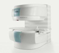
From Siemens Medical Systems;
A new, powerful, compact player in MRI. For both, patients and health care professionals, the mid-field has realized a giant step to cost efficient quality care. Obese patients and people with claustrophobia appreciate the comfortable side loading. The smallest pole diameter - 137 cm (54 inches) allows for optimal patient comfort.
Device Information and Specification
CLINICAL APPLICATION
Whole body
SE, FLASH, FISP, IR, FIR, STIR, TrueIR/FISP, FSE, MT, SS-FSE, MT-SE, MTC, MSE, EPI, PSIF
IMAGING MODES
Single, multislice, volume study, multi angle, multi oblique
512 x 512 full screen display
41 cm vertical gap distance
| | | |
• View the DATABASE results for 'MAGNETOM C™' (2).
| | | | | | Further Reading: | Basics:
|
|
| |
| | | | |
| | | |
|
| |
 | Look
Ups |
| |


