Magnetic Resonance - Technology Information Portal Welcome to MRI Technology
Info
  Sheets

Out-
      side


 'Echo' 
SEARCH FOR    
 
  2 3 5 A B C D E F G H I J K L M N O P Q R S T U V W X Y Z
Result : Searchterm 'Echo' found in 100 terms [] and 162 definitions []
previous     26 - 30 (of 262)     next
Result Pages : [1 2 3 4 5 6 7 8 9 10 11 12 13 14 15 16 17 18 19 20]  [ ... ]
Searchterm 'Echo' was also found in the following services: 
spacer
News  (7)  Resources  (10)  Forum  (35)  
 
Spin Echo SequenceInfoSheet: - Sequences - 
Intro, 
Overview, 
Types of, 
etc.MRI Resource Directory:
 - Sequences -
 
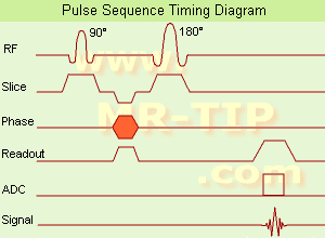
Spin Echo Timing Diagram (SE) The most common pulse sequence used in MR imaging is based of the detection of a spin or Hahn echo. It uses 90° radio frequency pulses to excite the magnetization and one or more 180° pulses to refocus the spins to generate signal echoes named spin echoes (SE).
In the pulse sequence timing diagram, the simplest form of a spin echo sequence is illustrated.
The 90° excitation pulse rotates the longitudinal magnetization (Mz) into the xy-plane and the dephasing of the transverse magnetization (Mxy) starts.
The following application of a 180° refocusing pulse (rotates the magnetization in the x-plane) generates signal echoes. The purpose of the 180° pulse is to rephase the spins, causing them to regain coherence and thereby to recover transverse magnetization, producing a spin echo.
The recovery of the z-magnetization occurs with the T1 relaxation time and typically at a much slower rate than the T2-decay, because in general T1 is greater than T2 for living tissues and is in the range of 100-2000 ms.
The SE pulse sequence was devised in the early days of NMR days by Carr and Purcell and exists now in many forms: the multi echo pulse sequence using single or multislice acquisition, the fast spin echo (FSE/TSE) pulse sequence, echo planar imaging (EPI) pulse sequence and the gradient and spin echo (GRASE) pulse sequence;; all are basically spin echo sequences.
In the simplest form of SE imaging, the pulse sequence has to be repeated as many times as the image has lines.
Contrast values:
PD weighted: Short TE (20 ms) and long TR.
T1 weighted: Short TE (10-20 ms) and short TR (300-600 ms)
T2 weighted: Long TE (greater than 60 ms) and long TR (greater than 1600 ms)
With spin echo imaging no T2* occurs, caused by the 180° refocusing pulse. For this reason, spin echo sequences are more robust against e.g., susceptibility artifacts than gradient echo sequences.

See also Pulse Sequence Timing Diagram to find a description of the components.
 
Images, Movies, Sliders:
 Shoulder Coronal T1 SE  Open this link in a new window
    

Courtesy of  Robert R. Edelman
 Shoulder Axial T1 SE  Open this link in a new window
 MRI Orbita T1  Open this link in a new window
    
 
spacer
 
• Related Searches:
    • Echo Time
    • T1 Weighted
    • Brain MRI
    • T1 Time
    • Pulse Sequence Timing Diagram
 
Further Reading:
  Basics:
Fast Spin Echo(.pdf)
Tuesday, 24 January 2006   by www.81bones.net    
Magnetic resonance imaging
   by www.scholarpedia.org    
FUNDAMENTALS OF MRI: Part I
   by www.e-radiography.net    
  News & More:
New MR sequence helps radiologists more accurately evaluate abnormalities of the uterus and ovaries
Thursday, 23 April 2009   by www.eurekalert.org    
MRI techniques improve pulmonary embolism detection
Monday, 19 March 2012   by medicalxpress.com    
Searchterm 'Echo' was also found in the following services: 
spacer
Radiology  (4) Open this link in a new windowUltrasound  (159) Open this link in a new window
Variable Echo Time
 
The variation of echo time from one RF pulse to the next.
spacer
MRI Resources 
Research Labs - Crystallography - Online Books - Journals - Used and Refurbished MRI Equipment - Patient Information
 
Asymmetrical Echoes
 
The echo time TE vary from echo to echo in the echo train.
spacer

• View the DATABASE results for 'Asymmetrical Echoes' (2).Open this link in a new window

Searchterm 'Echo' was also found in the following services: 
spacer
News  (7)  Resources  (10)  Forum  (35)  
 
Echo Planar ImagingInfoSheet: - Sequences - 
Intro, 
Overview, 
Types of, 
etc.MRI Resource Directory:
 - Sequences -
 
Echo Planar Imaging Timing Diagram (EPI) Echo planar imaging is one of the early magnetic resonance imaging sequences (also known as Intascan), used in applications like diffusion, perfusion, and functional magnetic resonance imaging. Other sequences acquire one k-space line at each phase encoding step. When the echo planar imaging acquisition strategy is used, the complete image is formed from a single data sample (all k-space lines are measured in one repetition time) of a gradient echo or spin echo sequence (see single shot technique) with an acquisition time of about 20 to 100 ms. The pulse sequence timing diagram illustrates an echo planar imaging sequence from spin echo type with eight echo train pulses. (See also Pulse Sequence Timing Diagram, for a description of the components.)
In case of a gradient echo based EPI sequence the initial part is very similar to a standard gradient echo sequence. By periodically fast reversing the readout or frequency encoding gradient, a train of echoes is generated.
EPI requires higher performance from the MRI scanner like much larger gradient amplitudes. The scan time is dependent on the spatial resolution required, the strength of the applied gradient fields and the time the machine needs to ramp the gradients.
In EPI, there is water fat shift in the phase encoding direction due to phase accumulations. To minimize water fat shift (WFS) in the phase direction fat suppression and a wide bandwidth (BW) are selected. On a typical EPI sequence, there is virtually no time at all for the flat top of the gradient waveform. The problem is solved by "ramp sampling" through most of the rise and fall time to improve image resolution.
The benefits of the fast imaging time are not without cost. EPI is relatively demanding on the scanner hardware, in particular on gradient strengths, gradient switching times, and receiver bandwidth. In addition, EPI is extremely sensitive to image artifacts and distortions.
spacer

• View the DATABASE results for 'Echo Planar Imaging' (19).Open this link in a new window


• View the NEWS results for 'Echo Planar Imaging' (1).Open this link in a new window.
 
Further Reading:
  Basics:
New Imaging Method Makes Brain Scans 7 Times Faster
Sunday, 9 January 2011   by www.dailytech.com    
Searchterm 'Echo' was also found in the following services: 
spacer
Radiology  (4) Open this link in a new windowUltrasound  (159) Open this link in a new window
Effective Echo Time
 
(TEeff) The contrast and the SNR of an MR image are determined primarily by the temporal position of the echo at which the phase encoding gradient has the smallest amplitude. The echo signal in this case undergoes minimal dephasing and has the strongest signal. The time period between the excitation pulse and this echo is the effective echo time.
spacer

• View the DATABASE results for 'Effective Echo Time' (2).Open this link in a new window

 
Further Reading:
  Basics:
RARE
Monday, 3 December 2012   by www2.warwick.ac.uk    
Clinical evaluation of a speed optimized T2 weighted fast spin echo sequence at 3.0 T using variable flip angle refocusing, half-Fourier acquisition and parallel imaging
Wednesday, 25 October 2006
Fast Spin Echo(.pdf)
Tuesday, 24 January 2006   by www.81bones.net    
MRI Resources 
Guidance - Lung Imaging - Artifacts - Absorption and Emission - MR Guided Interventions - Pathology
 
previous      26 - 30 (of 262)     next
Result Pages : [1 2 3 4 5 6 7 8 9 10 11 12 13 14 15 16 17 18 19 20]  [ ... ]
 Random Page
 
Share This Page
FacebookTwitterLinkedIn

MR-TIP    
Community   
User
Pass
Forgot your UserID/Password ?    


How AI will impact MRI :
only diagnostics 
saving time 
reducing cost 
makes planning obsolete 
reduce human knowledge 
not at all 

Look
      Ups


MR-TIP.com uses cookies! By browsing MR-TIP.com, you agree to our use of cookies.

Magnetic Resonance - Technology Information Portal
Member of SoftWays' Medical Imaging Group - MR-TIP • Radiology-TIP • Medical-Ultrasound-Imaging • 
Copyright © 2003 - 2024 SoftWays. All rights reserved. [ 26 April 2024]
Terms of Use | Privacy Policy | Advertising
 [last update: 2024-02-26 03:41:00]