 | Info
Sheets |
| | | | | | | | | | | | | | | | | | | | | | | | |
 | Out-
side |
| | | | |
|
| | | | |
Result : Searchterm 'Image Resolution' found in 1 term [ ] and 8 definitions [ ] and 8 definitions [ ], (+ 18 Boolean[ ], (+ 18 Boolean[ ] results ] results
| 1 - 5 (of 27) nextResult Pages :  [1] [1]  [2] [2]  [3 4 5 6] [3 4 5 6] |  | |  | Searchterm 'Image Resolution' was also found in the following services: | | | | |
|  |  |
| |
|
The image resolution is the level of detail of an image and a measurement of image quality. Higher resolution means more image detail, for example when two structures 1 mm apart are distinguishable in an image, this picture has a higher resolution than an image where they are not to distinguish.
More data points in an MR image (with same FOV) will decrease the pixel size, but not accurately improve the resolution because the different MRI sequences influence the contrast and the discernment of different tissues.
With high contrast and optimal signal to noise ratio, the image resolution is depend on FOV and number of data points of a picture, but T2* effects have an additional influence. | |  | | | | • Share the entry 'Image Resolution':    | | |
• View the NEWS results for 'Image Resolution' (1).
| | | | |  Further Reading: Further Reading: | | Basics:
|
|
News & More:
| |
| |
|  |  | Searchterm 'Image Resolution' was also found in the following service: | | | | |
|  |  |
| |
|
Inhomogeneity is the degree of lack of homogeneity, for example the fractional deviation of the local magnetic field from the average value of the field. Inhomogeneities of the static magnetic field, produced by the scanner as well as by object susceptibility, is unavoidable in MRI. The large value of gyromagnetic coefficient causes a significant frequency shift even for few parts per million field inhomogeneity, which in turn causes distortions in both geometry and intensity of the MR images.
Manufacturers try to make the magnetic field as homogeneous as possible, especially at the core of the scanner. Even with an ideal magnet, a little inhomogeneity is always left and is caused in addition by the susceptibility of the imaging object.
The geometrical distortion (displacement of the pixel locations) are important e.g., for some cases as stereotactic surgery. Displacements up to 3 to 5 mm have been reported. The second problem is the undesired changes in the intensity or brightness of pixels, which may cause problems in determining different tissues and reduce the maximum achievable image resolution.

Image Guidance
| |  | |
• View the DATABASE results for 'Inhomogeneity' (21).
| | | | |  Further Reading: Further Reading: | News & More:
|
|
| |
|  | |  |  | |  |  | Searchterm 'Image Resolution' was also found in the following services: | | | | |
|  |  |
| |
|
(DQA) This MRI scan or MRI procedure is used by system operators to verify system operation based on relevant image quality parameters like e.g., SNR, slice thickness, geometric distortion, slice position, image resolution and ghosting.
The quality assurance should carry out according to instructions of the manufacturer, normally using the head coil. In addition, SNR can be measured monthly on a selection of commonly used coils.
Weekly recording of these parameters is recommended for clinical MRI machines, as this allows early detecting of deviations from acceptable limits. | |  | |
• View the DATABASE results for 'Daily Quality Assurance' (3).
| | | | |  Further Reading: Further Reading: | News & More:
|
|
| |
|  |  | Searchterm 'Image Resolution' was also found in the following service: | | | | |
|  |  |
| |
|

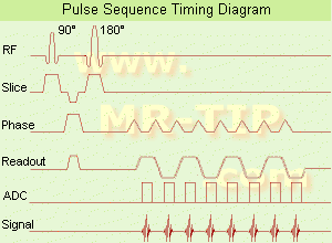
(EPI) Echo planar imaging is one of the early magnetic resonance imaging sequences (also known as Intascan), used in applications like diffusion, perfusion, and functional magnetic resonance imaging. Other sequences acquire one k-space line at each phase encoding step. When the echo planar imaging acquisition strategy is used, the complete image is formed from a single data sample (all k-space lines are measured in one repetition time) of a gradient echo or spin echo sequence (see single shot technique) with an acquisition time of about 20 to 100 ms.
The pulse sequence timing diagram illustrates an echo planar imaging sequence from spin echo type with eight echo train pulses. (See also Pulse Sequence Timing Diagram, for a description of the components.)
In case of a gradient echo based EPI sequence the initial part is very similar to a standard gradient echo sequence. By periodically fast reversing the readout or frequency encoding gradient, a train of echoes is generated.
EPI requires higher performance from the MRI scanner like much larger gradient amplitudes. The scan time is dependent on the spatial resolution required, the strength of the applied gradient fields and the time the machine needs to ramp the gradients.
In EPI, there is water fat shift in the phase encoding direction due to phase accumulations. To minimize water fat shift (WFS) in the phase direction fat suppression and a wide bandwidth (BW) are selected. On a typical EPI sequence, there is virtually no time at all for the flat top of the gradient waveform. The problem is solved by "ramp sampling" through most of the rise and fall time to improve image resolution.
The benefits of the fast imaging time are not without cost. EPI is relatively demanding on the scanner hardware, in particular on gradient strengths, gradient switching times, and receiver bandwidth. In addition, EPI is extremely sensitive to image artifacts and distortions. | |  | |
• View the DATABASE results for 'Echo Planar Imaging' (19).
| | |
• View the NEWS results for 'Echo Planar Imaging' (1).
| | | | |  Further Reading: Further Reading: | Basics:
|
|
| |
|  | |  |  |
|  | 1 - 5 (of 27) nextResult Pages :  [1] [1]  [2] [2]  [3 4 5 6] [3 4 5 6] |
| |
|
| |
 | Look
Ups |
| |