Magnetic Resonance - Technology Information Portal Welcome to MRI Technology
Info
  Sheets

Out-
      side


 'Array Coil' 
SEARCH FOR    
 
  2 3 5 A B C D E F G H I J K L M N O P Q R S T U V W X Y Z
Result : Searchterm 'Array Coil' found in 4 terms [] and 18 definitions []
previous     6 - 10 (of 22)     next
Result Pages : [1]  [2 3 4 5]
Searchterm 'Array Coil' was also found in the following services: 
spacer
Resources  (3)  Forum  (1)  
 
MAGNETOM C™InfoSheet: - Devices -
Intro, 
Types of Magnets, 
Overview, 
etc.MRI Resource Directory:
 - Devices -
 
www.medical.siemens.com From Siemens Medical Systems;
A new, powerful, compact player in MRI. For both, patients and health care professionals, the mid-field has realized a giant step to cost efficient quality care. Obese patients and people with claustrophobia appreciate the comfortable side loading. The smallest pole diameter - 137 cm (54 inches) allows for optimal patient comfort.
Device Information and Specification
CLINICAL APPLICATION
Whole body
CONFIGURATION
C-shaped open MRI
Multi channel imaging, CP Head//Neck Array Coil, Body/Spine Array Coil, large, Transmit Coil
SYNCHRONIZATION
ECG/peripheral: Optional/yes, respiratory gating
PULSE SEQUENCES
SE, FLASH, FISP, IR, FIR, STIR, TrueIR/FISP, FSE, MT, SS-FSE, MT-SE, MTC, MSE, EPI, PSIF
IMAGING MODES
Single, multislice, volume study, multi angle, multi oblique
TR
Min. TR 2.81 ms
TE
Min. TE 0.98 ms
512 x 512 full screen display
MEASURING MATRIX
64 x 64 to 512 x 512
FOV
0.5 - 40 cm
MAGNET TYPE
Permanent
BORE DIAMETER
or W x H
41 cm vertical gap distance
MAGNET WEIGHT
16000 kg
H*W*D
233 x 206 x 160 cm
STRENGTH
24 mT/m
5-GAUSS FRINGE FIELD
2.2 m / 2.2 m
Passive and active
spacer
 
• Related Searches:
    • MRI Equipment
    • Hardware
    • Device
    • Open MRI
    • Permanent Magnet
 
Further Reading:
  Basics:
Section 2: 510(k) Summary, MAGNETOM C! System Classification Name: Magnetic Resonance Diagnostic(.pdf)
   by www.accessdata.fda.gov    
MRI Resources 
Claustrophobia - Functional MRI - Cochlear Implant - Implant and Prosthesis pool - Mobile MRI Rental - Quality Advice
 
Simultaneous Acquisition of Spatial Harmonics
 
(SMASH) Several lines of data are acquired for each phase encoding step, which is also referred to as a k-space trajectory.
SMASH imaging with a four-element array coil is four times faster and can be used to achieve almost real-time imaging. The maximum reduction in acquisition time is determined by the number of array coil elements. Thus, the heart can be scanned with higher temporal resolution and increased spatial resolution.
SMASH and SENSE differ from other techniques in which only one line of k-space data is acquired for each phase encoding gradient step.

See Sensitivity encoding.
spacer

• View the DATABASE results for 'Simultaneous Acquisition of Spatial Harmonics' (3).Open this link in a new window

MRI Resources 
Online Books - Stimulator pool - Musculoskeletal and Joint MRI - Education pool - MRI Technician and Technologist Schools - RIS
 
AIRIS II™InfoSheet: - Devices -
Intro, 
Types of Magnets, 
Overview, 
etc.MRI Resource Directory:
 - Devices -
 
www.hitachimed.com/products/airis_2.asp 
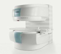
From Hitachi Medical Systems America Inc.;
the AIRIS II, an entry in the diagnostic category of open MR systems, was designed by Hitachi Medical Systems America Inc. (Twinsburg, OH, USA) and Hitachi Medical Corp. (Tokyo) and is manufactured by the Tokyo branch. A 0.3 T field-strength magnet and phased array coils deliver high image quality without the need for a tunnel-type high-field system, thereby significantly improving patient comfort not only for claustrophobic patients.
Device Information and Specification
CLINICAL APPLICATION
Whole body
CONFIGURATION
Vertical Field, Open MRI
QD Head, MA Head and Neck, QD C-Spine, MA or QD Shoulder, MA CTL Spine, QD Knee, Neck, QD TMJ, QD Breast, QD Flex Body (4 sizes), Small and Large Extrem., QD Wrist, MA Foot and Ankle (WIP), PVA (WIP)
SYNCHRONIZATION
Cardiac gating, ECG/peripheral, respiratory gating (2 modes)
PULSE SEQUENCES
SE, GE, GR, IR, FIR, STIR, FSE, ss-FSE, FLAIR, EPI -DWI, SE-EPI, ms - EPI, SSP, MTC, SARGE, RSSG, TRSG, MRCP, Angiography: CE, 2D/3D TOF
IMAGING MODES
Single, multislice, volume study
TR
SE: 30 - 10,000msec GE: 20 - 10,000msec IR: 50 - 16,700msec FSE: 200 - 16,7000msec
TE
SE : 10 - 250msec IR: 10 -250msec GE: 5 - 50 msec FSE: 15 - 2,000
SINGLE/MULTI SLICE
0.05 sec/image (256 x 256)
FOV
5cm to 42 cm continuous
2D: 2 - 100 mm; 3D: 0.5 - 5 mm
1280 x 1024
MEASURING MATRIX
512 x 512
PIXEL INTENSITY
Level Range: -2,000 to +4,000
Sub millimeter
MAGNET TYPE
Self-shielded, permanent
BORE DIAMETER
or W x H
110 x 43 cm
MAGNET WEIGHT
15,700 kg
H*W*D
79 x 111 x 73 cm
POWER REQUIREMENTS
208/220/240 V, single phase
COOLING SYSTEM TYPE
Air-cooled
STRENGTH
15 mT/m
2.0 m lateral, 2.5 m vert./long
Auto shimming, 3-axis/patient, and volume shim
spacer

• View the DATABASE results for 'AIRIS II™' (2).Open this link in a new window

Searchterm 'Array Coil' was also found in the following services: 
spacer
Resources  (3)  Forum  (1)  
 
ARTOSCAN™ - MInfoSheet: - Devices -
Intro, 
Types of Magnets, 
Overview, 
etc.MRI Resource Directory:
 - Devices -
 
www.gemedicalsystems.com/rad/mri/products/artoscan/specs.html Developed by GE Lunar; the ARTOSCAN™-M is designed specifically for in-office musculoskeletal imaging. ARTOSCAN-M's compact, modular design allows placing within a clinical environment, bringing MRI to the patient. Patients remain outside the magnet at all times during the examinations, enabling constant patient-technologist contact. ARTOSCAN-M requires no special RF room, magnetic shielding, special power supply or air conditioning.
The C-SCAN™ (also known as Artoscan C) is developed from the ARTOSCAN™ - M, with a new computer platform.
Device Information and Specification
CLINICAL APPLICATION
Dedicated extremity
CONFIGURATION
Closed
Linear and dual phased array coil, knee, ankle, wrist coils
PULSE SEQUENCES
SE, GE, IR, STIR, FSE, 3D CE, GE-STIR, 3D GE, ME, TME, HSE
IMAGING MODES
Single, multislice, volume study, fast scan, multi slab, cine
TR
12 - 5,000 msec
TE
5 - 220 msec
SINGLE SLICE
0.8 sec/image
MULTISLICE
0.8 sec/image
FOV
10 - 20 cm
SLICE THICKNESS
2D: 2 mm - 10 mm;
3D: 0.6 mm - 10 mm
512 x 512
MEASURING MATRIX
256 x 256 maximum
PIXEL INTENSITY
4,096 gray lvls, 256 lvls in 3D
MAGNET TYPE
Ferrite Permanent
MAGNET WEIGHT
965 kg
POWER REQUIREMENTS
100/110/200/220/230/240V
STRENGTH
10 mT/m
5 GAUSS FRINGE FIELD, radial/axial
28 cm/60 cm
Passive
spacer

• View the DATABASE results for 'ARTOSCAN™ - M' (3).Open this link in a new window

MRI Resources 
Artifacts - Hospitals - Safety Products - Veterinary MRI - Software - Examinations
 
Coil DiameterInfoSheet: - Coils - 
Intro, 
Overview, 
etc.
 
MRI coils with a small diameter obtain a higher signal to noise ratio (SNR) than coils with a large diameter. A surface coil with a small diameter can be used to improve the resolution because the area of interest is around the optimal signal depth. The field of view of a (superficial) surface coil is half the diameter of the coil. A disadvantage is a lower sensitive volume of the coil. By combining several coils with small diameters (phased array coil) to record the signal simultaneously and independently, the SNR level improves considerably.
spacer

• View the DATABASE results for 'Coil Diameter' (3).Open this link in a new window

MRI Resources 
Portals - Equipment - Case Studies - Pediatric and Fetal MRI - Education - Breast MRI
 
previous      6 - 10 (of 22)     next
Result Pages : [1]  [2 3 4 5]
 Random Page
 
Share This Page
FacebookTwitterLinkedIn

MR-TIP    
Community   
User
Pass
Forgot your UserID/Password ?    


Personalized protocols (age, gender, body habitus, etc.) lead to :
more automated planning 
improved patient comfort 
better diagnostics 
optimized image quality 
nothing 

Look
      Ups


MR-TIP.com uses cookies! By browsing MR-TIP.com, you agree to our use of cookies.

Magnetic Resonance - Technology Information Portal
Member of SoftWays' Medical Imaging Group - MR-TIP • Radiology-TIP • Medical-Ultrasound-Imaging • 
Copyright © 2003 - 2024 SoftWays. All rights reserved. [ 25 April 2024]
Terms of Use | Privacy Policy | Advertising
 [last update: 2024-02-26 03:41:00]