 | Info
Sheets |
| | | | | | | | | | | | | | | | | | | | | | | | |
 | Out-
side |
| | | | |
|
| | | | |
Result : Searchterm '3 Dimensional Imaging' found in 1 term [ ] and 4 definitions [ ] and 4 definitions [ ], (+ 11 Boolean[ ], (+ 11 Boolean[ ] results ] results
| | previous 6 - 10 (of 16) nextResult Pages : [1] [2 3 4] |  | |  | Searchterm '3 Dimensional Imaging' was also found in the following service: | | | | |
| |  |
| |
|
•
In the 1930's, Isidor Isaac Rabi (Columbia University) succeeded in detecting and measuring single states of rotation of atoms and molecules, and in determining the mechanical and magnetic moments of the nuclei.
•
Felix Bloch (Stanford University) and Edward Purcell (Harvard University) developed instruments, which could measure the magnetic resonance in bulk material such as liquids and solids. (Both honored with the Nobel Prize for Physics in 1952.) [The birth of the NMR spectroscopy]
•
In the early 70's, Raymond Damadian (State University of New York) demonstrated with his NMR device, that there are different T1 relaxation times between normal and abnormal tissues of the same type, as well as between different types of normal tissues.
•
In 197 3, Paul Lauterbur (State University of New York) described a new imaging technique that he termed Zeugmatography. By utilizing gradients in the magnetic field, this technique was able to produce a two- dimensional image (back-projection). (Through analysis of the characteristics of the emitted radio waves, their origin could be determined.) Peter Mansfield further developed the utilization of gradients in the magnetic field and the mathematically analysis of these signals for a more useful imaging technique. (Paul C Lauterbur and Peter Mansfield were awarded with the 200 3 Nobel Prize in Medicine.)
•
1977/78: First images could be presented.
A cross section through a finger by Peter Mansfield and Andrew A. Maudsley.
Peter Mansfield also could present the first image through the abdomen.
•
In 1977, Raymond Damadian completed (after 7 years) the first MR scanner (Indomitable). In 1978, he founded the FONAR Corporation, which manufactured the first commercial MRI scanner in 1980. Fonar went public in 1981.
•
1981: Schering submitted a patent application for Gd-DTPA dimeglumine.
•
1982: The first 'magnetization-transfer' imaging by Robert N. Muller.
•
In 198 3, Toshiba obtained approval from the Ministry of Health and Welfare in Japan for the first commercial MRI system.
•
1986: Jürgen Hennig, A. Nauerth, and Hartmut Friedburg (University of Freiburg) introduced RARE (rapid acquisition with relaxation enhancement) imaging. Axel Haase, Jens Frahm, Dieter Matthaei, Wolfgang Haenicke, and Dietmar K. Merboldt (Max-Planck-Institute, Göttingen) developed the FLASH ( fast low angle shot) sequence.
•
1988: Schering's MAGNEVIST gets its first approval by the FDA.
•
In 1991, fMRI was developed independently by the University of Minnesota's Center for Magnetic Resonance Research (CMRR) and Massachusetts General Hospital's (MGH) MR Center.
•
From 1992 to 1997 Fonar was paid for the infringement of it's patents from 'nearly every one of its competitors in the MRI industry including giant multi-nationals as Toshiba, Siemens, Shimadzu, Philips and GE'.
| | | | | | | | | | | | |  Further Reading: Further Reading: | | Basics:
|
|
News & More:
| |
| |
| | | | | |
| |
|

From GE Healthcare;
'EXCITE technology has the potential to open the door to new imaging techniques and clinical applications, leaping beyond conventional two and three- dimensional MRI to true 4D imaging that will improve the diagnosis of disease throughout the human body from head to foot.' Robert R. Edelman, M.D., Professor of Radiology at Northwestern University Medical School and Chairman, Department of Radiology, at Evanston Northwestern Healthcare.
Device Information and Specification CLINICAL APPLICATION Whole body Head and body coil standard; all other coils optional; open architecture makes system compatible with a wide selection of coils Optional 2D/ 3D brain and prostate Standard: SE, IR, 2D/ 3D GRE and SPGR, Angiography: 2D/ 3D TOF, 2D/ 3D Phase Contrast;; 2D/ 3D FSE, 2D/ 3D FGRE and FSPGR, SSFP, FLAIR, EPI, optional: 2D/ 3D Fiesta, FGRET, Spiral, TensorTR 1.3 to 12000 msec in increments of 1 msec TE 0.4 to 2000 msec in increments of 1 msec 2D 0.7 mm to 20 mm; 3D 0.1 mm to 5 mm 128x512 steps 32 phase encode 0.08 mm; 0.02 mm optional POWER REQUIREMENTS 480 or 380/415 less than 0.0 3 L/hr liquid heliumSTRENGTH SmartSpeed 23 mT/m, HiSpeed Plus 33 mT/m, EchoSpeed Plus 33 mT/m 4.0 m x 2.8 m axial x radial | | | |
• View the DATABASE results for 'Signa Infinity 1.5T™ with Excite' (2).
| | | | |
| | | | | |
| |
|
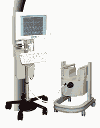
From MagneVu;
The MagneVu 1000 is a compact, robust, and portable, permanent magnet MRI system and operates without special shielding or costly site preparation.
This MRI device utilizes a patented non-homogeneous magnetic field image acquisition method to achieve high performance imaging. The MagneVu 1000 MRI scanner is designed for MRI of the extremities with the current specialty areas in diabetes and rheumatoid arthritis. Easy access is afforded for claustrophobic, pediatric, or limited mobility patients. In August 1998
FDA marketing clearance and other regulatory approvals have been received. Until 2008, over 1 30 devices in the US are in use. Some further developments of MagneVu's extremity scanner are: 'truly Plug n' Play MRI™' and iSiS ( which adds wireless capability to the second generation MV1000-XL).
Device Information and Specification IMAGING MODES 3-dimensional multi-echo data acquisition | | | |
• View the DATABASE results for 'MagneVu 1000' (3).
| | | | | | Further Reading: | News & More:
|
|
| |
| | | Searchterm '3 Dimensional Imaging' was also found in the following service: | | | | |
| | |
| |
|
( BOLD) In MRI the changes in blood oxygenation level are visible. Oxyhaemoglobin (the principal haemoglobin in arterial blood) has no substantial magnetic properties, but deoxyhaemoglobin (present in the draining veins after the oxygen has been unloaded in the tissues) is strongly paramagnetic. It can thus serve as an intrinsic paramagnetic contrast agent in appropriately performed brain MRI. The concentration and relaxation properties of deoxyhaemoglobin make it a susceptibility , e.g. T2 relaxation effective contrast agent with little effect on T1 relaxation.
During activation of the brain, the oxygen consumption of the local tissue increase by approximately 5% with that the oxygen tension will decrease. As a consequence, after a short period of time vasodilatation occurs, resulting in a local increase of blood volume and flow by 20 - 40%. The incommensurate change in local blood flow and oxygen extraction increases the local oxygen level.
By using T2 weighted gradient echo EPI sequences, which are highly susceptibility sensitive and fast enough to capture the three- dimensional nature of activated brain areas will show an increase in signal intensity as oxyhaemoglobin is diamagnetic and deoxyhaemoglobin is paramagnetic. Other MR pulse sequences, such as spoiled gradient echo pulse sequences are also used.
As the effects are subtle and of the order of 2% in 1.5 T MR imaging, sophisticated methodology, paradigms and data analysis techniques have to be used to consistently demonstrate the effect.
As the BOLD effect is due to the deoxygenated blood in the draining veins, the spatial localization of the region where there is increased blood flow resulting in decreased oxygen extraction is not as precisely defined as the morphological features in MRI. Rather there is a physiological blurring, and is estimated that the linear dimensions of the physiological spatial resolution of the BOLD phenomenon are around 3 mm at best. | | | |
• View the DATABASE results for 'Blood Oxygenation Level Dependent Contrast' (6).
| | | | | | Further Reading: | | Basics:
|
|
News & More:
| |
| |
| | | | | |
| |
|
(DTI) Diffusion tensor imaging is the more sophisticated form of DWI, which allows for the determination of directionality as well as the magnitude of water diffusion. This kind of MR imaging can estimates damage to nerve fibers that connect the area of the brain affected by the stroke to brain regions that are distant from it, and can be used to determine the effectiveness of stroke prevention medications.
DTI (FiberTrak) enables to visualize white matter fibers in the brain and can map ( trace image) subtle changes in the white matter associated with diseases such as multiple sclerosis and epilepsy, as well as assessing diseases where the brain's wiring is abnormal, such as schizophrenia.
The fractional anisotropy (FA) gives information about the shape of
the diffusion tensor at each voxel. The FA is based on the normalized
variance of the eigenvalues. The fractional anisotropy reflects differences between an isotropic diffusion and a linear diffusion. The FA range is between 0 and 1 (0 = isotropic diffusion, 1 = highly directional).
The development of new imaging methods and some useful analysis techniques, such as 3- dimensional anisotropy contrast ( 3DAC) and spatial tracking of the diffusion tensor tractography (DTT), are currently under study. | | | |
• View the DATABASE results for 'Diffusion Tensor Imaging' (9).
| | |
• View the NEWS results for 'Diffusion Tensor Imaging' (2).
| | | | | | Further Reading: | Basics:
|
|
News & More:
| |
| |
| | | | |
| | | |
|
| |
 | Look
Ups |
| |


